Researchers from the U.S. Army are using PET scans of mice to study brain inflammation and gather more information about the progression of disease due to the Zika virus, according to a study published online September 12 in Molecular Imaging and Biology.
PET's highly sensitive molecular imaging capabilities make the modality a valuable tool for visualizing a variety of biological processes in live animal models, according to senior author Thomas Bocan, PhD, and colleagues from the U.S. Army Medical Research Institute of Infectious Diseases (USAMRIID).
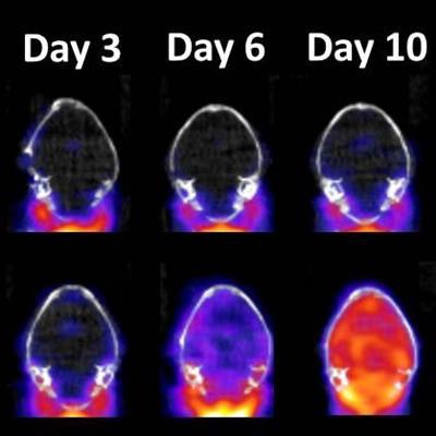
In the study, the USAMRIID researchers combined PET with a radiopharmaceutical probe known as DPA-714 to detect and quantify neuroinflammation in mice infected with the Zika virus. They found that levels of Zika virus in the mouse brain increased from the third day to the 10th day after infection. During that time, the mice also showed a two- to sixfold increase in global brain neuroinflammation based on F-18 DPA-714 PET imaging.
 Representative DPA-714-PET images (bottom row) of mouse brain at third, sixth, and 10th day after infection with Zika virus. Red area indicates neuroinflammation. Top row shows control images for comparison. Image courtesy of USAMRIID.
The findings demonstrate for the first time the ability of DPA-714-PET to detect and quantify Zika virus-related neuroinflammation disseminated throughout the brain in infected mice, according to the authors.
"Traditional methods of infectious disease research using animal models have provided limited information about disease progression until the study's end point, when investigators could analyze tissues from those animals," Bocan said in a statement from the institute. "Imaging studies allow us to gather enhanced information through longitudinal studies of the same animal during the course of the infection."
Noninvasive imaging with PET and other modalities can also reduce the number of animals in a study by permitting the use of animals as their own controls, the researchers added. In addition, therapeutic agents can be developed and tested using imaging for validation.
"The future is bright for the application of imaging in infectious diseases," Bocan said. "Measures of virus and bacteria distribution and the consequences of infection can be assessed in real-time in the same subject. In addition, treatment with countermeasures can be evaluated with a better understanding of the state of disease progression."


