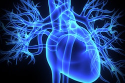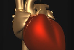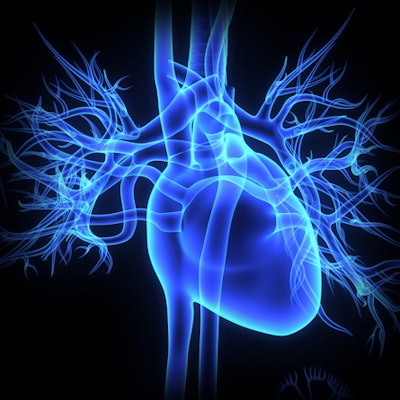
Researchers from Italy have created low-cost, individually tailored 3D-printed aortas that could work well for transcatheter aortic valve replacement (TAVR), according to an article published online May 26 in the Journal of Cardiovascular Computed Tomography.
Prior research has highlighted the various benefits of using patient-specific 3D-printed models of the aortic root to plan complex procedures such as TAVR or implantation, as well as help predict potential TAVR leaks.
Aiming to evaluate the feasibility of planning TAVR with 3D-printed aortas, co-first author Dr. Marco Gatti and colleagues from the University of Turin examined the 3D-printed aortas of 20 patients who underwent aortic valve replacement at the hospital between January and October 2017.
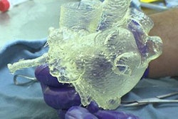
They tasked one radiologist and one radiologic technologist -- both trained in the creation of 3D files -- with the construction of a separate 3D-printed aorta for each of the patients based on their contrast-enhanced CT scans.
To make the model, the radiologist and technologist individually segmented the CT scans using computer software (OsiriX MD 9.0, Pixmeo), converted the segmented scans into stereolithography (STL) format, and refined the 3D model using open source software (Meshmixer 3.4, Autodesk). Finally, they 3D printed the models with an ultraflexible filament (NinjaFlex, NinjaTek) in a 3D printer (Ultimaker 2 Extended+, Ultimaker).
There was no significant difference between the annulus diameters of the 3D-printed aortas created by the radiologist and technologist, the group found. In addition, the measurements of the 3D-printed models displayed good agreement with measurements taken from the reference cardiac CT scans preoperatively, as well as from the actual aortas during aortic valve replacement surgery.
On average, the diameters of the 3D-printed aortas had a 96% correlation with the reference CT scans and a 95% correlation with the intraoperative measurements.
What's more, production of the 3D-printed models was relatively quick and cost-effective. It took approximately 10 to 15 minutes to segment and process the 3D model, 60 minutes to 3D print it, and less than five minutes to postprocess it after printing. The team was able to 3D print 10 models in just under 10 hours using printing materials that cost roughly $1.17 per model.
"This approach may offer a reliable, not expensive, patient-specific preoperative planning opportunity," the authors wrote. "It provides a unique interactive platform to the final user with both visual and tactile experiences, which are critical for simulation of the procedure but not available in imaging data per se."
The researchers noted that the study's low sample size as well as measuring only the aortic annulus during diastole may have limited the scope of their findings.
"With future optimizations, including development of printers and materials that better reflect the mechanical properties of the aortic annulus and associated calcifications, this developing technology may acquire an important role in the preprocedural workup [for TAVR]," they wrote.