Researchers from Canada have developed a new technique that uses real-time imaging of 3D CT models to guide chest wall tumor resection. The method was more accurate and easier to use than conventional thoracoscopy, according to an article recently published online in the Annals of Thoracic Surgery.
Several studies have shown that minimally invasive surgery for chest wall tumors can improve patient outcomes compared with open chest wall tumor resection, yet its adoption in clinical practice has been slow due to the limited visibility of the procedure, noted lead author Dr. Kazuhiro Yasufuku, PhD, and colleagues from University Health Network in Toronto.
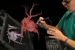
Seeking to increase the procedure's viability, the researchers collaborated with scientists from the Guided Therapeutics (GTx) surgery program at the health network to develop a proprietary surgical navigation technique that combines traditional video-based thoracoscopy with conebeam CT and optical tracking to perform augmented real-time image guidance (Ann Thorac Surg, August 16, 2018).
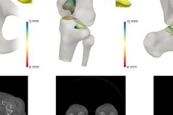
To develop the technique, the group created and scanned a chest wall phantom with conebeam CT and then reconstructed the scans into 3D models. Next, the investigators contoured the tumors and their surgical margins in these models using open-source segmentation software (ITK-Snap), and they set up real-time tracking of the phantom with custom 3D visualization software (GTx Eyes, GTx).
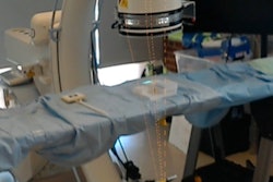
Finally, they attached infrared reflective markers to the phantom and the thoracoscope, which allowed clinicians to look at 3D models of the chest wall and its tumors on 2D displays from the perspective of the surgical tool. Once the new surgical navigation platform was ready, the group had 15 thoracic surgeons use it to locate tumors and their corresponding surgical margins in the chest wall phantom.
Using the new image-guidance method reduced localization errors for tumors in the medial and superior regions of the chest to a statistically significant degree, as well as improved the accuracy of measuring the surgical margins, compared with standard thoracoscopy.
What's more, the surgeons claimed that using the technique helped ease their workload -- considerably reducing the mental and physical demand, frustration levels, and effort required to complete the task on a subjective scale of zero (low workload) to 20 (high workload).
| Conventional thoracoscopy vs. thoracoscopy with augmented reality | ||
| Conventional thoracoscopy | Thoracoscopic navigation with augmented reality | |
| Localization error for medial tumor | ~16.5 mm | ~2.5 mm |
| Localization error for superior surgical margin | ~10 mm | ~1.5 mm |
| Surgeon performance workload | 10.1 | 4.4 |
| Surgeon frustration | 13 | 5.4 |
In follow-up questionnaires, the surgeons agreed that the new navigation method helped with the identification of 110 out of 120 (92%) tumors and margins. They also felt that the technique boosted their confidence when identifying the tumors and margins.
"Our thoracoscopic surgical navigation system improved tumor and surgical margin localization accuracy during minimally invasive chest wall tumor surgery with a phantom model," the authors wrote. "This system also provided surgeons with augmented real-time imaging, which increased their performance and confidence, while decreasing their frustration."