Researchers from Pennsylvania have created a patient-specific 3D-printed head phantom that may facilitate the testing of new protocols on 7-tesla MRI scanners.
The group, led by doctoral candidate Sossena Wood from the University of Pittsburgh, began by acquiring a 3-tesla MRI dataset of a man. Next, the researchers segmented the scans and separated them into eight compartments, each representing a distinct type of tissue within the head, including the brain, eyes, air cavities, and muscle, among others.
They then converted the dataset into a 3D model using computer-aided design software (Geomagic Studio, Solid Technologies) and printed the model as five separate parts with a 3D printer.
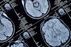
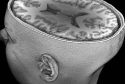
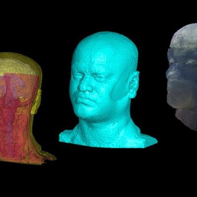
 A 3T MRI dataset (left), segmented 3D model (middle), and 3D-printed phantom head (right). Image courtesy of the University of Pittsburgh Radiofrequency Research Facility.
To make the 3D-printed head phantom as realistic as possible, they used a plastic material that allowed for highly detailed structures, and they also deposited liquids into the model to resemble fluids in the head such as cerebrospinal fluid.
Wood and colleagues plan to use the 3D-printed phantom head for various applications, from testing the safe use of certain brain implants in 7-tesla MRI scanners to detecting changes to tissue temperature based on shifts in radiofrequency.
"We wanted to develop an anthropomorphic phantom head to help us better understand these issues [with 7-tesla MRI] by providing a safer way to test the imaging," adviser Tamer Ibrahim, PhD, said in a statement from the university. "We [can] use the device to analyze, evaluate, and calibrate the MRI systems and instrumentation before testing new protocols on human subjects."