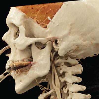
Radiologists from Massachusetts identified ear calcification on the cinematically rendered CT scans of a patient with traumatic brain injury -- effectively allowing them to uncover a rare diagnosis without performing a physical exam, according to an article published online January 28 in Endocrine.
Auricular calcification, or calcium buildup in the ear, is an elusive condition often associated with endocrine pathologies such as diabetes and adrenal insufficiency. Traditionally, patients would undergo radiography to confirm the presence of auricular calcification only after it was suspected on a physical exam, but incidental detection on CT has been more common due to the asymptomatic nature of the condition, noted Dr. Michael Caton and Dr. Fiona Malone from Brigham and Women's Hospital.
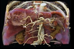
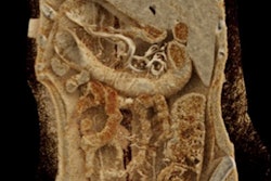
In a recent case study, Caton and Malone acquired the CT scans of a 47-year-old man with a history of traumatic brain injury who had previously undergone craniectomy to relieve pressure in the skull. They used advanced volume rendering software (syngo.via, Siemens Healthineers) to cinematically render the CT data and generate photorealistic images.
The resulting cinematically rendered images greatly improved depth perception as well as the visualization of surface textures and underlying calcification below the surface of the skin, compared with conventional CT, Caton told AuntMinnie.com. As a result, the researchers were able to spot calcification in both of the patient's ears, which physicians had not recognized on a prior physical exam.
 Head CT scan of a 47-year-old man with a history of traumatic brain injury (A). Auricular calcification is evident on the patient's cinematically rendered CT scans (B, C). Image courtesy of Dr. Michael Caton.
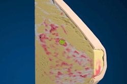
Manually adjusting the CT window and level settings also gave the researchers the opportunity to selectively emphasize or suppress tissues with different radiographic densities on the cinematically rendered images -- enabling them to alternate between views of the skin's surface and the underlying bony and soft-tissue structures.
"This [technique] enhances our understanding of how pathology is seen by our referring colleagues and is helpful when making recommendations to referring physicians; we can be more specific than just saying 'correlate clinically,' " he said. "For radiologists, clinicoradiographic correlation with cinematic rendering gives us an opportunity to perform a virtual physical exam."
Furthermore, the highly detailed information available on cinematically rendered images may help strengthen the relationship between radiologists and physicians of other specialties, as they work together to create patient-specific models to aid treatment planning, according to the researchers. The improved visualization that cinematic rendering provides could also benefit patient care in several other ways, including patient education.
"I hope that cinematic rendering can demystify medical imaging for our patients," Caton said. "My experience sharing cinematically rendered images with patients has been overwhelmingly positive; patients (and nonradiology physicians) find such images to be very intuitive. I think this could greatly improve the recognition and visibility of our work as radiologists to our patients."