German researchers have developed a new PET imaging technique that shows early promise in predicting Alzheimer's disease progression in mice, according to a preclinical study published in the April issue of the Journal of Nuclear Medicine.
The PET scans target the 18-kD translocator protein (TSPO), which is highly expressed in activated microglia, making it an effective biomarker in the assessment of inflammation in the brain. By imaging microglial activation levels in mice, the researchers were able to determine the activity that influenced cognitive outcomes in an amyloid mouse model. The accumulation of amyloid plaques is associated with the onset of Alzheimer's disease (J Nucl Med, April 2019, Vol. 60:4, pp. 548-554).
For this study, researchers acquired PET images for 10 transgenic mice with beta-amyloid proteins and seven wild-type mice. TSPO-PET imaging of activated microglia was performed at eight, 9.5, 11.5 and 13 months, while beta-amyloid PET scans were done at eight and 13 months.
After imaging, the researchers conducted memory tests on the mice in a water maze to measure the average travel time from the start to a platform for each day of training, and to calculate the distance traveled at the last day of training. After completing the water maze task, immunohistochemistry analyses were performed for microglia, amyloid, and synaptic density.
Transgenic mice with the highest TSPO-PET signal in the forebrain or other regions associated with spatial learning tended to have better cognitive performance in the water maze, while beta-amyloid signals in the same areas of the brain showed no correlation to cognitive outcomes in the maze. In fact, transgenic mice with higher TSPO expression at eight months had much better cognitive outcomes in the water maze and higher synaptic density as confirmed by immunochemistry analyses.
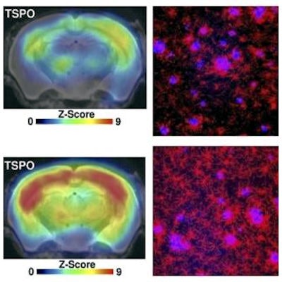
![Images show the multimodal correlation analysis of cognitive testing with terminal PET and immunohistochemical results in mice at the end of the study. Representative PET images (z score on MRI template), immunohistochemistry (fused methoxy-X04 [blue] and Iba1 [red]), and white matter findings of individual mice show either low (orange) or high (magenta) markers of microglial activation at study termination. Images courtesy of Focke et al and J Nucl Med.](https://img.auntminnie.com/files/base/smg/all/image/2019/04/am.2019_04_05_16_02_5671_TSPO_PET_Alzheimers_JNM.png?auto=format%2Ccompress&dpr=2&fit=max&q=70&w=700) Images show the multimodal correlation analysis of cognitive testing with terminal PET and immunohistochemical results in mice at the end of the study. Representative PET images (z score on MRI template), immunohistochemistry (fused methoxy-X04 [blue] and Iba1 [red]), and white matter findings of individual mice show either low (orange) or high (magenta) markers of microglial activation at study termination. Images courtesy of Focke et al and J Nucl Med.
Images show the multimodal correlation analysis of cognitive testing with terminal PET and immunohistochemical results in mice at the end of the study. Representative PET images (z score on MRI template), immunohistochemistry (fused methoxy-X04 [blue] and Iba1 [red]), and white matter findings of individual mice show either low (orange) or high (magenta) markers of microglial activation at study termination. Images courtesy of Focke et al and J Nucl Med."This study provides the first evidence that the level of microglial activation could be a far better predictor of current and future cognitive performance than beta-amyloid levels," said co-author Dr. Matthias Brendel, of Ludwig Maximilian University of Munich, in a statement. "Keeping the limitations of mouse models in mind, it could be crucial to modify an individual's microglial activation state to ameliorate future cognitive decline. We believe that a balanced microglia activation is crucial for prevention of cognitive impairment."