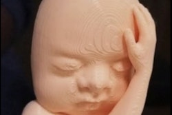
Researchers from Saudi Arabia have created 3D-printed models based on the CT scans of children with craniofacial deformities. Examining the models significantly increased the parents' likelihood of consenting to surgical treatment, according to a study recently published online in the Journal of Craniofacial Surgery.
The group, led by Dr. Feras Alshomer from King Khalid University Hospital in Riyadh, investigated the potential utility of 3D-printed models to aid consultations with parents of children who have craniofacial deformities that require surgical correction.
For complex conditions such as craniosynostosis, providing sufficient information about the patient's condition represents a crucial step in the management of these patients, especially since surgical treatment has functional and cosmetic implications.
Clinicians typically refer to the CT scans of patients in order to explain craniofacial deformities to their parents. Yet most parents find it difficult to understand their child's abnormality following this approach, and parents always express the need for additional information before choosing which surgical option they prefer, the authors noted.
To facilitate family counseling for patients with craniosynostosis, Alshomer and colleagues obtained the CT scans of seven children with craniofacial deformities and made individually tailored 3D-printed models for each case. Creation of models involved extracting and rendering 3D virtual models using open-source software (3D Slicer), converting 3D models into files suitable for printing using a different open-source software application (Cura, Ultimaker), and finally generating 3D-printed skulls with a desktop 3D printer (Ultimaker 2+, Ultimaker). It required approximately 26 hours and $5.20 in material costs to create each 3D-printed model (J Craniofac Surg, March 28, 2019).
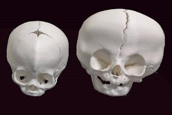
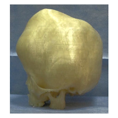
 3D virtual skull model based on patient CT scans (left). 3D-printed craniofacial model (right). Images courtesy of Dr. Feras Alshomer.
The researchers held individual consultations with the parents of each of the seven patients, during which they presented both conventional 3D CT scans and patient-specific 3D-printed models.
On follow-up questionnaires, the parents claimed to have a much clearer grasp of the pathology and were less likely to need more information about the condition and surgical intervention after examining 3D-printed skulls, compared with conventional 3D CT scans (p = 0.05; p = 0.019). In addition, examining 3D-printed models in place of 3D CT scans helped the parents more readily come to a decision regarding the proposed surgical intervention (p = 0.028).
However, both methods provided the parents with an equally clear view of how the skull might look after surgery. And the parents seemed to understand potential complications associated with the condition better by looking at 3D CT scans than at 3D-printed models.
Relying on 3D-printed skull models for surgical consultations offers an easy-to-use, affordable modality of education for parents of children with craniofacial deformities -- helping the parents decide on the appropriate surgical intervention without requiring additional information, Alshomer told AuntMinnie.com.
"I do believe that with the recent advancements in this technology, every physician should know and learn how to have their own 3D printing facility, without the need to outsource patient information and rely on extremely expensive printing facilities to deliver such tools," he said.