Since its debut in the 1980s, cardiac MRI has become an important clinical tool in helping to diagnose and treat heart disease. It is considered the gold standard in diagnostics because it is noninvasive and produces high-quality images that enable clinicians to visualize and quantify blood flow in the heart, aorta, and large vessels.
Despite these advantages, cardiac MRI is often a last resort in imaging modalities, primarily because image acquisition is difficult. Patients must remain very still and hold their breath multiple times. Some patients may need anesthesia to endure the test, increasing the risk. Plus, it takes longer to perform than other diagnostics, requiring a highly skilled technologist and radiologist, and the results may not be immediately available. These challenges may also reduce the usability of cardiac MRIs in emergent situations.
Other limitations to routine clinical application of cardiac MRI include availability of the hardware, cost, and perhaps most important, physician and technologist education. While some institutions already using cardiac MRI are challenged by the increasing demand, other institutions are unable to adopt the technology due to lack of skilled technologists to acquire the examination and clinicians to process and interpret the images.
Applying 4D flow technology
Advances in imaging technology have not only provided better quality studies but also relieved many of the difficulties with administering it. With earlier 2D-based systems, physicians had to determine ahead of time the necessary flow measurements and assist technologists in acquiring each sequence at the appropriate location. Each flow measurement required a separate acquisition, often requiring multiple breath holds.
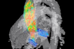
The introduction of 4D flow MRI -- 3D flow imaging over time -- is eliminating many of these challenges. Making its way out of research facilities and into clinical practice, 4D flow MRI enables clinicians to conduct comprehensive hemodynamic flow assessments, providing valuable assessment of the 3D and multidirectional nature of intracardiac blood flow.
These capabilities have been game changers for my practice. With 4D flow, we can image from the top of the arch through the apex of the heart, obtaining coverage through the entire heart and great vessels. It requires less planning time because we acquire the entire volume set; in fact, I no longer even need to be present.
With 4D flow, we also have been able to simplify our protocols. While not everything can be obtained in 4D flow, the information from the 4D flow sequence enables us to decrease some of the other sequences we were acquiring. In addition, the ease with which the 4D flow sequence is acquired enables other technologists with less cardiac training to run the sequence. Our facility is lucky to have dedicated cardiac technologists who are excellent, but that is not always the case in other practices. The simplified protocols mean any MRI technologist can successfully capture the necessary data.
Patients also see numerous benefits from 4D flow. It does not require patients to hold their breath, which opens up the possibilities of imaging younger patients without anesthesia and sicker patients who are typically unable to meet the breath-holding demands. We are now able to image patients as young as 6 years old without anesthesia. For patients with congenital heart diseases who often need to be imaged frequently, reducing the number of times they receive anesthesia is a significant benefit to their long-term health.
Perhaps most significant is the decreased amount of time necessary to scan a patient and analyze the images. Time slots for cardiac MRI scans in our practice were previously 90 minutes. With 4D flow, we are now able to perform these studies in 45-minute time slots. Additionally, I can now do all of my postprocessing after the patient has left, with no concern that I didn't capture the necessary images.
Gaining insights and speed through AI
Cardiac MRI isn't just time-consuming to capture but also to analyze. The labor-intensive nature makes it an ideal candidate for an application of artificial intelligence (AI). Medical imaging is a core part of patient care, and embedding it with AI can have a huge impact. For cardiac MRI, specifically, use of 4D flow generates exponentially more data. To help us effectively address the increasing quantity of information, we leverage a cloud-based AI platform. This platform offers the scalability and compute power needed to analyze massive amounts of 4D flow images and give us real-time, interactive insights to speed diagnosis.
AI also helps us more quickly analyze 2D function images, which are preprocessed prior to opening the study. The process of drawing circles and analyzing the 2D images, which used to take 20 to 40 minutes, has decreased dramatically. Now I spend five minutes looking at the contours and making a few adjustments, saving significant time and reducing my workload considerably.
While some practices may see barriers to introducing 4D flow and AI, these technologies are ultimately worth the time and the effort. 4D flow, combined with AI analytics, makes the cardiac MRI process easier for patients and more efficient for healthcare providers, and it provides the best diagnostic information to treat the patient.
Dr. Melany B. Atkins is a diagnostic radiologist with the Fairfax Radiological Consultants in Fairfax, VA.
The comments and observations expressed are those of the author and do not necessarily reflect the opinions of AuntMinnie.com.