Examining 3D-printed models based on head CT scans enabled clinicians to customize implants required for eye socket fracture repair and reduce the time to complete the surgery by roughly 50% in a new study, recently published online in Academic Radiology.
The group from the University of Michigan turned to 3D printing technology to help overcome the challenge of precisely sizing and positioning bioabsorbable implants needed to protect sensitive regions of the eye during orbital fracture repair.
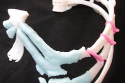
 Sterilizable 3D-printed models of the orbital bone with a fracture (left) and after repair (right). Image courtesy of Dr. William Weadock.
Standard practice for orbital bone fracture repair involves shaping an initially flat implant to fit the curvature of the orbital floor via trial and error. However, this approach is subject to improper shaping and placement of implants, which occasionally results in conditions such as enophthalmos, or posterior displacement of the eye, lead author Dr. William Weadock and colleagues noted.
Seeking to improve upon current methods, the researchers acquired the CT scans of several patients scheduled to undergo orbital fracture repair and used these data to create individually tailored 3D-printed models of the orbital bone for each of the patients (Acad Radiol, August 27, 2019).
The protocol for creating the models included segmenting the CT scans, converting the files into stereolithography (STL) format, and exporting them as virtual 3D models into 3D modeling software (Meshixer, Autodesk). During processing, the researchers superimposed the virtual 3D models of the nonfractured orbital bone onto those of the fractured orbital bone to fill in the fracture gap.
The final step was to print out the combined orbital bones with a 3D printer (Form 2, Formlabs). Manufacturing and sterilization of the 3D-printed models took approximately eight hours, including five hours just for 3D printing.
With a personalized 3D-printed model in hand, the surgeons were able to visualize the extent of the eye socket fracture and then shape the bioabsorbable implant to the contours of the fractured orbital bone. Follow-up CT scans later confirmed the proper positioning of the implant.
Weadock and colleagues applied this technique to the surgical repair of fractured eye socket bones caused by trauma in three patients.
Overall, they found that relying on 3D-printed models reduced the time required to complete surgery from a hospital average of 93.3 minutes to 48.3 minutes -- in turn, minimizing the patients' risk of postoperative edema, among other complications.
"The nearly 50% decrease in operating room time has potential implications for patient safety, with decreased anesthesia time," the authors wrote. "Additionally, decreased operating time will also result in decreased institutional costs."