Chest CT accurately detected the novel coronavirus disease (COVID-19) in the vast majority of high-risk individuals prior to confirmation by laboratory testing, though it also misidentified COVID-19 for the adenovirus in a small percentage of cases in a new study, published online March 4 in the American Journal of Roentgenology.
For the study, Dr. Yan Li and Dr. Liming Xia examined data from 53 patients evaluated for suspicion of COVID-19 at Tongji Hospital in Wuhan, China, the original site of the outbreak. The patients underwent chest CT exams and nucleic acid testing with reverse transcription polymerase chain reaction (RT-PCR). Their average age was 58 years, and roughly half were women.
The radiologists who examined the CT scans of these patients detected COVID-19 in 96.1% of all cases eventually confirmed by RT-PCR. They were able to differentiate COVID-19 from common bacterial infections based on CT alone in all but two cases.
Of note, CT showed abnormal patterns suggestive of COVID-19 in two other cases that DNA testing later revealed to be adenovirus infection and not COVID-19. Still, CT overall proved more effective than laboratory testing at early detection of COVID-19, confirming the disease an average of three days before laboratory testing in 69.8% of the patients.
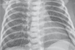
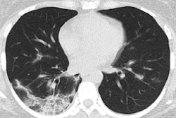
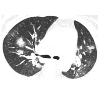
 CT image of a 58-year-old woman with adenovirus infection showing ill-defined patchy ground-glass opacities and segmental and subpleural consolidations in both lungs, with most lesions along bronchovascular bundles. Image courtesy of the AJR.
"It is valuable for radiologists to recognize that the CT findings of COVID-19 overlap with the CT findings of diseases caused by viruses from a different family, such as adenovirus, and have differences as well as similarities with viruses within the same family," Li and Xia wrote.
Among the patient cohort, the most common CT findings were ground-glass opacities and consolidation with or without vascular enlargement (96.1% of patients), followed by interlobular septal thickening (70.6%), air bronchogram sign (68.6%), and air trapping (11.8%). Most of these findings were similar to CT features previously reported in patients with severe acute respiratory syndrome (SARS) and Middle East respiratory syndrome (MERS).
The researchers did identify CT findings that distinguished COVID-19 from SARS and MERS in several cases: the presence of a reversed halo sign in 3.9% of patients and pulmonary nodules with a halo sign in 17.6%. In addition, COVID-19 patients more frequently had multifocal involvement on CT, compared with unifocal involvement for SARS and MERS.
The group also found that about 20% of initial CT scans showed fibrosis and striplike lesions with deformation of the bronchus, indicating that COVID-19 lesions were likely present on CT before external symptoms appeared and that CT should have been performed earlier in these cases.
"We are confident that CT has a high accuracy and may be useful as a standard method for the diagnosis of COVID-19," the authors concluded. "The use of CT for the diagnosis of viral pneumonia allows patients with suspected [coronavirus] infection to be isolated and treated in time for recovery, thus optimizing patient management."