Imaging, specifically MRI and CT, helped clinicians determine that a mass located on the chin of a newborn girl was a second oral cavity, according to a recent case report. The cavity contained unerupted teeth and an accessory tongue that moved in sync with her oral tongue.
The case of diprosopus, an extremely rare congenital disorder in which all or parts of the face are duplicated on the head, is one of about 35 that have been reported since 1900. These patients should be monitored closely, and imaging should be used to help identify and devise treatment plans, noted the authors of the report (BMJ Case Reports, May 19, 2020).
"In the case of suspected craniofacial duplication, associated syndromes should be ruled out and appropriate imaging employed to determine the extent of involvement of adjacent structures, which will ultimately guide surgical planning," wrote Alexandra Hamberis, of the Medical University of South Carolina (MUSC) College of Medicine, and colleagues.
Isolated oral and mandible duplication without associated syndromes has been reported only a handful of times, with the first case in 1948. In that case, a 2-day-old child was discovered to have a right mandibular mass with a unilateral duplication of the mouth and tongue with a blind pouch and synchronous accessory tongue movement, which almost mirrors the circumstances in the current case report.
Craniofacial duplications occur more often in females, and they often are associated with conditions such as cleft lip and palate, Klippel-Feil syndrome, and Pierre Robin sequence. The current case is extremely rare because the girl's partial craniofacial duplication is an isolated anomaly, with no associated syndromes or abnormalities, the authors wrote.
The 3rd trimester
Prenatal imaging during the third trimester of pregnancy showed that the girl had a right mandibular mass. After she was born at 40 weeks and four days to a healthy mother via an uncomplicated vaginal delivery, the girl was referred to otolaryngology to have the mass examined, according to the authors.
There was no family history of facial malformations, and the child wasn't exposed to any agents known to cause abnormalities in utero. The mass caused no respiratory or feeding issues, they wrote.
A physical exam revealed a 1- to 2-cm mass along her right mandible with intraoral displacement of the girl's tongue to the left. There was a small sinus tract, located 1 cm inferior and lateral to the right oral commissure. The approximately 13-mm deep sinus tract was adjacent to the mass but had no connection to her oral cavity.
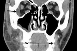
A closer look revealed that the girl's mandibular alveolar ridge was widened by several millimeters on the left, and she couldn't depress her right lower lip. An MRI performed shortly after her birth showed an approximately 2.5 x 2 x 2-cm lesion arising from the right mandibular body. Several unerupted teeth also were detected, the authors noted.
The doctors found nothing else remarkable, so she remained in the hospital nursery until she was discharged.
When the child was brought back to the hospital for her two-week checkup, she was found to be healthy. She had no feeding or oral complications and was gaining weight. The external area of the mass had a raw surface at times. Also, a small tongue protruded from the mass and appeared to move in conjunction with her oral tongue while she fed from a bottle.
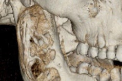
A CT of the mandible taken during that visit revealed an ossified soft-tissue mass of the right mandibular body containing teeth. Soft tissue was present along the surface of the bony area that extended caudally to the skin surface below the right side of the chin. No aggressive osseous features were revealed, and there was no evidence that injury or other stimuli formed new bone, the authors wrote.
Surgical removal
When the girl was 6 months old, surgery was performed to remove the duplicated mandible, provide bony contouring of the mandible, and close the soft-tissue defect with adjacent tissue.
Doctors resected and traced her mucosal lining and associated salivary glands to the mandible. The lining that extended to the mandible and appeared as mucosa overlying the alveolar arch was peeled off. Several primary teeth that faced toward the duplicate oral cavity were found in the underlying bone. Accessory teeth were pulled, sockets were drilled down to contour the mandible, and any remaining dental tissue was removed, they wrote.
On pathology, the removed mass was found to contain benign squamous mucosa, a salivary gland, cortical bone, skeletal muscle, and dental pulp with six benign molar teeth.
By a six-month follow-up visit, the girl's incisions had healed. She was feeding without difficulty but had persistent inability to depress her right lower lip, which may be due to her oral depressor muscles not being stimulated or failing to develop during embryonic development, Hamberis and colleagues wrote.
Be prepared
When cases like this are identified in utero, priority should be given to securing the airway when it is time for delivery, the group explained. Also, an otolaryngology team should be present during delivery. Management of craniofacial duplications depends on the patient's symptoms.
If the abnormality doesn't cause breathing or feeding problems, then surgery may be delayed for up to three years to get a better view of the patient's teeth, the authors wrote.
In addition to using CT, MRI, or both for surgical preparation, imaging should be used to differentiate accessory teeth from unerupted permanent teeth.
"Premature intervention may lead to removal of tooth buds that are a part of the normal mandibular anatomy," they wrote.


_20210915221258.png?auto=format%2Ccompress&fit=crop&h=167&q=70&w=250)