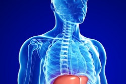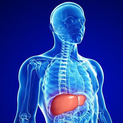
Contrast-enhanced ultrasound (CEUS) is a promising tool to diagnose some forms of liver disease, according to a recent review in Hepatoma Research.
Dr. Stephanie Wilson and Dr. Christina Merrill from Calgary, Canada, reviewed the unique benefits of CEUS over contrast-enhanced CT and MRI scans for liver imaging. In their September 1 report, they wrote that CEUS has the advantages of dynamic real-time imaging, capability for subtraction imaging, and being a purely intravascular contrast agent, which prevents leaking outside of tumors.
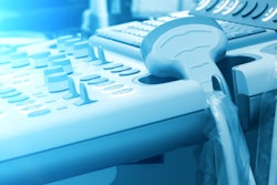
"CEUS provides exquisite vessel resolution and is a completely noninvasive imaging option that produces the same information as CT and MR, which have historically been used to diagnose focal liver lesions," stated Wilson in a press release from the International Contrast Ultrasound Society. "In addition, ultrasound is portable and may be brought to a patient's bedside, so it is often less disruptive for a patient."
The prevention of leaking is a crucial advantage over CT and MRI, which have a well-recognized interstitial phase, according to the authors. It also allows clinicians to see contrast microbubbles in all phases of imaging.
The differences between CEUS and CT and MRI may be particularly prominent in the portal venous phase, the authors noted. In this phase, CT and MRI often show pseudoenhancement from interstitial contrast, while CEUS will more accurately show washout in malignant tumors.
Finally, the authors explained that CEUS has specific software to aid with subtraction. This allows clinicians to obtain high-quality images in thin septations and small nodules, the authors noted.