Dual-energy CT (DECT) offers new ways to improve diagnosis of disease, and the technology has continued to improve over the past decade. But radiologists must be aware of DECT's potential pitfalls -- particularly artifacts -- in order to use it effectively, according to an overview published February 19 in RadioGraphics.
Understanding why DECT produces artifacts and how to avoid them is crucial to the successful use of the technology, wrote a team led by Dr. Anushri Parakh of Massachusetts General Hospital in Boston.
"The ability of radiologists and technologists to anticipate [the occurrence of artifacts] and provide recommendations for proper selection of patients, intravenous and oral contrast media, and scan acquisition parameters is key to obtaining good-quality DECT images," it wrote.
Parakh's group offered a primer of DECT technology, outlining its types and a description of potential pitfalls.
Types of DECT. DECT currently falls into two categories: Source-based or detector-based. Source-based DECT includes the following:
- Dual-source DECT (two x-ray tubes/detectors arranged perpendicular to each other)
- DECT with rapid tube voltage switching (peak voltage of the x-ray tube alternates between high and low energies)
- Split-filter DECT, in which the filter is composed of gold and tin and splits the x-ray into two beams, one high energy and one low energy
- Dual-spin DECT (patient undergoes two consecutive image acquisitions at different tube voltages)
Detector based DECT includes the following:
- Dual-layer DECT (single x-ray tube and two layers of detectors, with the top layer absorbing low-energy photons and bottom layer absorbing high-energy photons)
- Photon-counting DECT (still in research phase: a semiconductor detector converts x-ray photons into electrical signals)
Pitfalls related to image acquisition, processing, and display. Radiologists should take the following into consideration when performing DECT exams:
- Patient selection. Optimize DECT protocols by taking patient body weight and transverse diameter into consideration. "In patients with large body habitus, DECT may yield higher image noise and reduced image quality due to photon starvation," the team noted.
- Contrast medium selection. The appearance of iodine contrast appearance can be amplified on DECT, appearing as streaks on the image; reducing the iodine concentration in the contrast medium may be needed.
- Patient positioning. "Patients may need to be purposely off-centered to ensure that the organ of interest lies completely within the spectral field of view," the group cautioned.
- Temporal misregistration. This can affect image quality and lead to "spectral skew," in which data from the two energies being used are offset from each other.
- Image processing. Carefully select kernel, decomposition ratio, and thresholding parameters, the team wrote.
- Image display. Wider window settings are often needed to view DECT images.
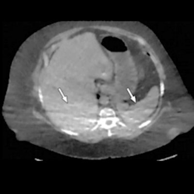
 Photon starvation in a large patient. Axial portovenous-phase abdominopelvic CT was performed in a 242-lb patient using rapid kilovolt peak (kVp) switching DECT. (a) Material density iodine image shows dark bands of artifact (arrows) across the liver and spleen. (b) On an MD-water image, the streaks appear as bright bands (arrows). (c) On a 65 kilo-electron volt (keV) image, the artifacts are much less apparent (arrows). (d) On a 140 kVp-equivalent image, the artifacts are not visualized. Images and caption courtesy of the RSNA.
There's no doubt that DECT is a promising technology, but radiologists must understand it to use it effectively, according to the team.
"DECT is an exciting innovation in CT technology, with tremendous capabilities for improving diagnosis and adding value to patient care in routine clinical practice," the group concluded. "[Yet] inherent in this technology are certain artifacts that need to be recognized to realize the full value of DECT and avoid diagnostic errors."