Researchers from China have developed a radiomics model capable of identifying on CT scans various immunohistochemical (IHC) characteristics associated with thyroid cancer. The model could bolster the noninvasive evaluation of thyroid nodules, according to an article recently published online in the American Journal of Roentgenology.
Typical visual evaluation of thyroid nodules on CT is often unable to distinguish benign from malignant nodules, requiring clinicians to perform a biopsy and subsequent immunohistochemical analysis for confirmation. However, there is potential to leverage texture analysis techniques to uncover immunohistochemical information noninvasively, principal investigator Jiabing Gu and colleagues from the University of Jinan noted.
"When IHC information is hidden on CT images, it may be possible to discern the relation between this information and radiomics by use of texture analysis," Gu said in a statement from the American Roentgen Ray Society.
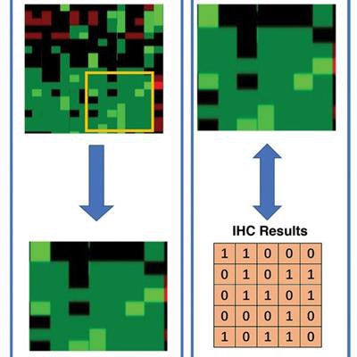
 Radiomics workflow for drawing immunohistochemical characteristics from CT scans. Image courtesy of AJR.
Exploring the utility of radiomics in thyroid cancer diagnosis, the researchers developed a radiomics model that used machine learning to examine 86 textural features on CT scans, with the aim of identifying immunohistochemical features associated with thyroid cancer (AJR, August 28, 2019).
To test the model, they acquired the CT scans of 103 patients suspected of having thyroid nodules who underwent surgery. They imported the scans into open-source computer software (3D Slicer) for image registration, segmentation, 3D modeling, and automated radiomics analysis. Gu and colleagues also externally validated the model on a separate dataset of 20 cases.
The model was able to identify the presence of various markers of thyroid nodules -- the proteins cytokeratin 19, galectin 3, and thyroid peroxidase -- by analyzing the CT scans, the group found. The model yielded accuracies ranging from 80% to 85% in the training and validation cohorts, as confirmed by actual immunohistochemical analysis of thyroid biopsies.
"This [radiomics] model may be used to identify benign and malignant thyroid nodules," especially for nodules that are difficult for radiologists to identify visually on CT, Gu said.