Ultrasound beats mammography as the best imaging modality for initial evaluation of breast cancer in symptomatic women ages 30 to 39, according to a new study published in the November issue of the American Journal of Roentgenology.
For women younger than 40, breast imaging is typically done to evaluate focal areas of concern, such as palpable lumps, researchers at the University of Washington in Seattle and the Seattle Cancer Care Alliance wrote. But these women are also those who tend to have dense breast tissue, which affects mammography's performance.
Because most lumps are not cancer, ultrasound and mammography help distinguish between women who need a biopsy and those who can safely be followed, said lead author Dr. Constance Lehman.
"Mammography is still our best tool for screening women 40 and older, but ultrasound is the modality of choice in evaluating symptomatic women under 40," Lehman told AuntMinnie.com.
The study cohort included all women 30 to 39 years of age who had ultrasound and mammography in response to focal breast signs or symptoms between January 2002 and August 2006. Lehman's team determined benign versus malignant outcomes by biopsy or imaging surveillance and through linkage with a tumor registry with a minimum 24-month follow-up.
The team also calculated overall cancer yield, sensitivity, specificity, negative predictive value (NPV), and positive predictive value (PPV) of ultrasound and mammography (AJR, November 2012, Vol. 199:5, pp. 1169-1177).
"To our knowledge, this study represents the largest analysis to date of breast ultrasound and adjunct mammography for the evaluation of women 30 to 39 years old who present with focal breast signs or symptoms," the group wrote. "Moreover, this study is the first to examine the most effective use of imaging for the diagnostic workup of this patient population and the only study to investigate this imaging application within the past decade."
The researchers identified 1,208 areas of concern in 954 patients, with benign outcomes in 1,185 of 1,208 individuals (98.1%) and malignant outcomes in 23 of 1,208 (1.9%). The group determined the following:
- Ultrasound's sensitivity in the patient population was 95.7%, while mammography's sensitivity was 60.9%.
- Mammography led in specificity at 94.4%, compared with 89.2% for ultrasound.
- NPV was 99.9% for ultrasound and 99.2% for mammography.
- PPV was 13.2% for ultrasound and 18.4% for mammography.
- Mammography identified one additional cancer in an asymptomatic area in a 32-year-old woman who was found to have a BRCA2 gene mutation.
"Our results show that breast ultrasound has 95.7% sensitivity for cancer detection at the site of focal breast concerns and 99.9% NPV, [which demonstrates its effectiveness] as a primary imaging modality for this patient cohort," the group wrote.
 |
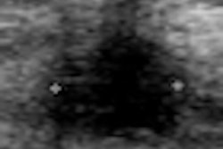
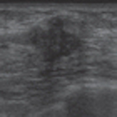
| 35-year-old woman who presented with palpable left breast lump. Whole-breast craniocaudal (above left) and mediolateral oblique (above right) and spot-magnification craniocaudal (below left) and mediolateral (below right) mammographic images show no abnormality at area of clinical concern, marked by BB. All images courtesy of the American Roentgen Ray Society. |
 |
 |
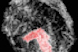
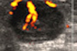
| Targeted ultrasound image above reveals solid mass with irregular shape and indistinct and angular margins. BI-RADS 5 assessment was made. Histopathology from ultrasound-guided core needle biopsy showed invasive ductal carcinoma. |
When in Rome ...
Current American College of Radiology (ACR) guidelines recommend using mammography as the primary imaging modality in women 30 years and older, while ultrasound is reserved for women younger than 30. This recommendation contrasts with European practice, Lehman said: In Europe, breast ultrasound is more commonly used as the first evaluation tool for symptomatic women younger than 40.
"The American College of Radiology and radiologists across the U.S. are trying to image wisely and limit radiation," Lehman told AuntMinnie.com. "Our study findings show that we could make the same shift our European colleagues have, and be more selective with mammography with symptomatic women under 40."
Although the cancer risk of radiation from mammography is still being debated, it's clear that patients should be exposed to radiation only if the benefits of the imaging test outweigh the potential and real risks, Lehman's team wrote. Additional years of unnecessary mammography may put women 30 to 39 years old at a small, but not insignificant, increased lifetime risk of radiation-induced breast cancer.
"Our results suggest that adjunct mammography may safely be reserved for particular high-risk cases, such as those patients with a highly suspicious lesion on ultrasound or those with a known gene mutation or strong family history," the team concluded.