Shear-wave elastography is sensitive and specific for detecting injuries to the Achilles tendon, and it can serve as a more objective method than other imaging modalities for diagnosis and treatment, according to research from the University of Wisconsin School of Medicine and Public Health.
In a small study, the researchers found that shear-wave imaging yielded 92% sensitivity and 83% specificity for diagnosing moderate to severe Achilles tendinopathy. They also found that injured Achilles tendons were thicker and had increased echogenicity and hyperemia.
"Overall, this study validates the possibility that shear-wave imaging can be a useful adjunct and is sensitive and specific for diagnosing moderate to severe Achilles tendinopathy," said Dr. Michael Llewellyn, PhD. He presented the results at the recent American Institute of Ultrasound in Medicine (AIUM) annual meeting in Orlando, FL.
Achilles tendinopathy
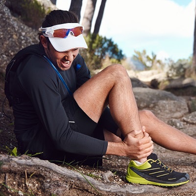
The largest and strongest tendon in the human body, the Achilles tendon is subjected to significant stress and strain during walking and running. As a result, Achilles tendon injuries represent a high percentage of all athletic injuries, and the prevalence increases significantly with age, Llewellyn said.
Achilles tendinopathy occurs due to acute or chronic microtears over time, and it can lead to increased pain, disability, and higher likelihood of a complete tear, he said. Diagnosis currently relies on a clinical exam as well as imaging studies -- most commonly ultrasound. Ultrasound findings associated with Achilles tendinopathy include increased thickening of the tendon, increased echogenicity, and increased hyperemia. However, imaging findings often don't correlate with patient symptoms.
"So a new method to add to our current repertoire of imaging is needed," he said.
Enter shear-wave imaging, an ultrasound elastography technique that uses focused ultrasound beams to produce a supersonic shear wave, which can be used to calculate the mechanical properties of tissue.
"It's not operator-dependent, unlike strain elastography," Llewellyn said.
While shear-wave imaging is already being used as an inexpensive and simple method for detecting cancer in other parts of the body, not much is known about how the modality acts in the musculoskeletal system, he said. As a result, the researchers set out to determine if shear-wave imaging could serve as an objective, operator-independent method for diagnosing moderate to severe Achilles tendinopathy.
As part of another prospective study underway at the institution, 12 subjects with unilateral, moderate to severe Achilles tendinopathy received shear-wave elastography on the Aixplorer ultrasound system (SuperSonic Imagine) with a high-frequency linear-array transducer. With the ankle in the neutral, relaxed position, the Achilles tendon was imaged in the longitudinal plane; B-mode, power Doppler, and aponeurosis shear-wave speed data were acquired on the midsubstance of the tendon. Imaging was performed on both the tendinopathic side and the contralateral tendon, which served as the control for the study.
Shear-wave imaging was used to assess tendon thickness, echogenicity, hyperemia, and shear-wave speed. The patients consisted of nine men and three women and ranged in age from 42 to 65.
Highly sensitive, specific
At a threshold shear-wave speed of 12 m/sec, shear-wave imaging produced 92% sensitivity and 83% specificity for diagnosing moderate to severe Achilles tendinopathy.
| Shear-wave imaging for Achilles tendinopathy | |||||
| Contralateral tendon | Tendinopathic tendon | ||||
| Average thickness | 6.6 mm | 10.3 mm | |||
| Hyperemia score (1-3) | 1.4 | 2.7 | |||
| Echogenicity score (1-3) | 1.4 | 2.6 | |||
| Shear-wave speed | 13.3 m/sec | 10.3 m/sec | |||
As expected, the tendinopathic side was significantly thicker than the contralateral side in the patients.
"However, what we didn't expect was that the control side was significantly thicker than you would have expected from a normal tendon, based on data that's been recorded in the literature," he said.
The tendinopathic tendons were also more hyperemic than the contralateral side. In addition, as expected, the tendinopathic side had increased echogenicity compared with the contralateral side, Llewellyn said. He noted, however, that the contralateral side had more hypoechogenicity than what has been reported in the literature.
The shear-wave speed on the tendinopathic side was lower than on the contralateral side. This was expected based on what has been reported in animal studies, he said.
All differences were statistically significant (p < 0.01).
Llewellyn acknowledged a number of limitations of the study, including the small number of patients. In addition, the tendons on the control side were not "normal," on average, compared with data reported in the literature. He also noted that elastography analysis is potentially sensitive to tendon tension during ankle dorsiflexion.
"We controlled for this, but the effect of tendon tension is very large," he said.


