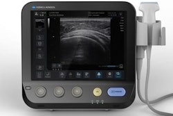
All-optical ultrasound technology can provide real-time 2D imaging of biological tissue with imaging capabilities comparable to those of an electronic high-frequency ultrasound system, and it could even be used safely with MRI, according to research published in the August issue of Biomedical Optics Express.
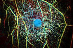
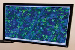
Researchers from University College London in the U.K. have developed a prototype of an all-optical ultrasound system using a spatially extended nanocomposite optical ultrasound generator, a fiber-optic acoustic receiver, and what they call "eccentric illumination." In testing on a deceased zebra fish as well as a pig artery manipulated to emulate the dynamics of pulsing blood, the system was capable of a sustained frame rate of 15 Hz, a dynamic range of 30 decibels, a penetration depth of 6 mm, and a resolution of 75 x 100 micrometers.
What's more, the system doesn't require electronic components in the imaging probe, enabling its use in the MRI environment and providing physicians with a more comprehensive view of an area of interest such as a tumor or blood vessel, according to the researchers.
"All-optical ultrasound imaging probes have the potential to revolutionize image-guided interventions," said Erwin Alles, PhD, in a statement. "A lack of electronics and the resulting MRI compatibility will allow for true multimodality image guidance, with probes that are potentially just a fraction of the cost of conventional electronic counterparts."
The device uses lightbeam scanning mirrors designed to improve image quality and acquire images in different modes. When using the system clinically, doctors could rapidly toggle between modes on a single instrument, according to the researchers (Biomed Opt Express, August 2018, Vol. 9:8, pp. 3481-3494).
While optical ultrasound systems have previously been shown to produce high-quality 2D and 3D images, image acquisition took hours -- too slow to be used in a clinical setting. However, the new prototype can acquire and display images at video rates, according to the group.
"Through the combination of a new imaging paradigm, new optical ultrasound generating materials, optimized ultrasound source geometries, and a highly sensitive fiber-optic ultrasound detector, we achieved image frame rates that were up to three orders of magnitude faster than the current state of the art," Alles said.
In an effort to adapt the technology for clinical use, the researchers are now working to develop a long, flexible imaging probe for free-hand operation. Miniaturized versions for endoscopic applications are also being developed.