What is a good response to deep brain stimulation (DBS) treatments for people with Parkinson's disease?
Researchers at the University of California, San Francisco (UCSF) and UC Berkeley are getting closer to understanding how DBS changes the medication burden for Parkinson's disease patients, according to a project presented May 13 at the International Society for Magnetic Resonance in Medicine (ISMRM) 2025 meeting.
The work may eventually enable healthcare providers to select patients for whom DBS is best suited.
"In an ideal paradigm, we can consider that DBS is working when the patient wouldn't need to take as much medication to treat their symptoms, so their medication burden would go down," stated Devin Schoen, a doctoral candidate working in the Morrison Neuromodulation Lab at UCSF.
DBS typically targets the subthalamic nucleus (STN) and the globus pallidus interna (GPi) brain regions to alleviate motor symptoms of Parkinson's disease. UCSF is one of the largest DBS centers in the world and houses more than a decade of retrospective data, Schoen noted.
However, "up to 30% of Parkinson's disease patients who get DBS don't actually see any meaningful motor improvement, and we know that there are a lot of factors that play into how someone will respond to the therapy," Schoen explained.
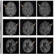
Toward a more personalized approach, Schoen and colleagues collected pre- and postoperative MRI imaging and clinical information for a large cohort for a study of this kind (n = 228) -- 110 STN patients and 128 GPi patients.
The team used Lead DBS, a free software specific to DBS studies, for image processing and reconstructing and localizing the electrodes. They stimulated the electric fields to find the volume of activated tissue (VAT) and then compared changes in the standard levodopa equivalent daily dose (LEDD) across patients and time points.
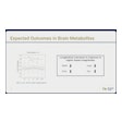
"We do know that LEDD is not the gold standard for DBS outcomes," Schoen said, "but it is a good option for us at this stage because we do have it for every single patient. It allows us to have this really large cohort, and there is precedent for using it in the field, [but] we are actively collecting the motor and cognitive outcomes for those patients who we're able to, and those will be folded into alter analysis."
In both STN and GPi-implanted patients, Schoen's team identified group-level aggregate centers of optimal stimulation and explored target- and hemisphere-specific differences for refining clinical targeting strategies. They determined the following:
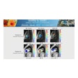
In the STN group, closer proximity to the aggregate center was correlated with greater LEDD reduction in the right hemisphere (r = -0.425, 95% CI [-0.639, -0.150], p = 0.004), suggesting that the aggregate center is an effective stimulation zone associated with reduced medication needs, the team reported. For the GPi group, the initial aggregate center based on medication reduction yielded no significant correlations with LEDD.
 Devin Schoen, ISMRM 2025
In the GPi cohort, a significant correlation was found between right-sided VAT overlap with the GPi target region and LEDD (r = 0.341, 95% CI [0.023, 0.595], p = 0.036), indicating that greater overlap is associated with larger reductions in LEDD. No other significant correlations were found between VAT overlap and LEDD.
 Devin Schoen, ISMRM 2025
"The precise location within the STN is more critical than overall volume of stimulation tissue for therapeutic effectiveness," the team noted. "The distinct responses between GPi and STN imply that DBS targets may require different stimulation strategies: optimizing for broader coverage in the GPi while focusing on specific sweet-spot regions in the STN."
Additionally, the aggregate centers based on medication changes provided insights into therapeutic zones; however, individual variability suggests a potential role for patient-specific imaging, according to the team.
Schoen referenced the team's preprint manuscript titled, "Boundary Complexity of (Sub-) Cortical Areas Predict Deep Brain Stimulation Outcomes in Parkinson’s Disease," for their full methods and results.
The group plans to identify a subset of GPi patients with brittle dyskinesias to investigate whether their VAT overlap or proximity to the aggregate center is associated with increased LEDD, indicating enhanced medication tolerance, or decreased LEDD, indicating reduced medication needs.
Check out AuntMinnie’s full coverage of ISMRM 2025 here.


