One optimum way to reduce inconclusive lung biopsy results and better detect malignancies is to use FDG-PET/CT for the image-guided procedure. Researchers from Brazil found that PET/CT had advantages over conventional CT guidance.
In a study published December 31, 2020, in the Journal of Nuclear Medicine, researchers prospectively evaluated more than 300 patients undergoing evaluation for suspected lung cancer and found that FDG-PET/CT-guided biopsies found a statistically significant greater percentage of malignant lesions than invasive procedures guided by CT alone.
The clinical benefit of PET/CT biopsy guidance is quicker and more appropriate treatment for patients with suspected lung cancer.
"Our results may influence the decision of the preferred method for guidance of percutaneous biopsy of lung lesions, if both PET/CT and CT are available," wrote the researchers, led by first author Dr. Juliano Julio Cerci, PhD, from Quantum Diagnostic Imaging in Curitiba, Brazil.
CT-guided biopsy has long been considered an "effective and safe method in the diagnostic workup in several different clinical settings," the researchers noted, and if FDG-PET's metabolic information were combined with CT's anatomical findings, the results "may optimize the diagnostic yield of image-guided interventions." A head-to-head comparison of the two modalities for percutaneous lung biopsies, however, has never been conducted, they added.
To fill that void, Cerci and colleagues analyzed results from 216 patients (mean age, 65.9 ± 12.1 years) who underwent FDG PET/CT-guided percutaneous biopsy and 125 patients (mean age, 65.4 ± 13.2 years) who underwent the same procedure using CT only as the guide. All 341 subjects underwent whole-body biopsies on the same PET/CT system (Discovery STE-16, GE Healthcare), which featured a CT fluoroscopic imaging system. They then were followed for a minimum of six months.
In the first round of biopsies, the FDG-PET/CT-guided approach was statistically superior (p = 0.001), with the discovery of 170 malignant lesions (79%), compared with 77 malignant lesions (62%) among the CT-guided biopsy group. The remaining 46 benign lung lesions (21%) in the FDG-PET/CT-guided group were significantly less than the 48 benign lesions (38%) among the CT-guided biopsy patients (p = 0.001).
FDG-PET/CT also recorded significantly fewer inconclusive results (p < 0.001) in the first round of biopsies. The hybrid-imaging approach reported only five inconclusive results (2%), compared with 13 inconclusive results (10%) with CT-guided biopsies. Follow-up biopsies showed no statistically significant difference between CT-guided procedures, which concluded there were 12 malignant lesions (92%), while FDG-PET/CT confirmed five malignant results (100%) from all five indeterminate cases.
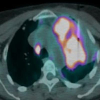
 Images are from a patient with lung mass who was referred for FDG-PET/CT. The PET image (A) shows multiple lesions in the left lung, lymph nodes, and bones. The axial CT thoracic image (B) shows the placement of the coaxial guide needle in the lesion that does not differentiate atelectasis from the metabolic active lung lesion. The axial PET/CT fused image (C) confirms the positioning of the coaxial guide needle in the metabolic border of the lesion, which ensures that metabolically active specimens will be collected with a needle that can be inserted 2 cm further. A pathological analysis confirmed adenocarcinoma. Images courtesy of the Journal of Nuclear Medicine.
"Once we integrate CT's excellent anatomical information, especially important to evaluate vessels and important structures related to the biopsy path, with the metabolic characterization provided by FDG-PET images, sampling from the hypermetabolic portion of the apparently larger morphological lesion seen on CT becomes feasible, which is more likely to yield representative material for microscopic analysis," Cerci and colleagues wrote.
The researchers also investigated how well patients tolerated the two biopsies procedures and found "similar complication rates in both groups, with no life-threatening adverse events or fatalities reported. Hence, given the size of our sample and the observed outcomes, we deem both methods quite safe overall."
As for the clinicians performing the tasks, their exposure to radiation was similar with FDG-PET/CT and CT alone, "since our approach did not require a repeat FDG injection during the biopsy procedure," they added.