Photon-counting CT (PCCT) not only improves image quality for arterial phase abdominal scans compared to conventional CT, but it also reduces radiation dose by almost 20%, according to research presented at the 2022 RSNA meeting.
The findings are good news for oncology patients, wrote a team led by presenter Dr. Dirk Graafen, PhD, of Johannes Gutenberg-Universität Mainz in Germany.
"Image quality improvement of photon-counting detector CT for arterial-phase abdominal scans holds the potential to increase diagnostic certainty of oncologic imaging ... [and] further dose reduction might be achievable," the group explained.
Clinicians seek to reduce patient exposure to radiation, especially on CT imaging, which can impart high doses. Photon-counting CT shows promise for having considerable impact on abdominal oncologic imaging when it comes to reducing radiation dose, Graafen's team noted.
"Photon-counting detector CT ... is expected to have a substantial impact on abdominal oncologic imaging," the group wrote.
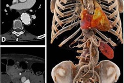
The investigators sought to compare photon-counting CT for abdominal imaging to conventional energy-integrating CT. They conducted a study that included 84 patients who underwent both types of exams for the abdomen in the arterial phase. On the qualitative side, the researchers compared the two scans for noise, contrast-to-noise of vessel, kidney cortex, and hypervascular liver lesions to liver parenchyma; on the quantitative side, they compared the two exams for radiation dose exposure. Three readers evaluated qualitative results using a five-point Likert scale.
Graafen and colleagues found that mean CT dose index-volume (CTDIvol) was 18% lower and mean effective dose 26% lower in PCCT scans than in conventional CT exams. Contrast-to-noise ratio of kidney cortex and vessel to liver parenchyma were higher at lower energies (less than or equal to 60 keV) on photon-counting CT compared to conventional CT (p < 0.004); lesion conspicuity was also better at low keV (p < 0.03), according to the investigators. An extra benefit? Interreader agreement among the three radiologists for photon-counting CT images was 0.80 (reference, 1).
"Photon-counting detector CT significantly improves image quality for arterial-phase oncologic abdominal imaging," Graafen concluded. "[Its] potential is the increase in diagnostic certainty of oncologic imaging and reduced radiation dose."
Beyond abdominal
There were a number of studies presented at the meeting that underscored PCCT's ability to reduce radiation dose across a variety of indications, not just abdominal imaging. A team from Duke University in Durham, NC, found that the technology improved image quality of pediatric chest exams compared with conventional CT -- without increasing radiation dose, and French investigators found that using photon-counting detector technology for chest CT angiographic exams also improved image quality -- and slashed radiation dose by more than 50%.
The results from the latter presentation were particularly dramatic: A group led by Dr. Martine Remy-Jardin, PhD, of the University Centre of Lille in France reported a 52% reduction in mean dose length product when comparing photon-counting CT to dual-source CT, at 172.6 mGy-cm versus 339.4 mGy-cm (p < 0.0001). Additionally, the investigators noted that photon-counting CT exam time was shorter compared to dual-source CT, at 0.93 seconds compared with four seconds.
"Photon-counting CT has the potential to improve perfusion imaging while maintaining morphologic image quality with a radiation dose reduced by 50%," this team concluded.