Photon-counting CT (PCCT) performs just as well as MRI for quantifying liver fat in obese patients and can enable opportunistic screening for fatty liver disease, according to research results delivered at the recent RSNA meeting in Chicago.
The findings suggest that CT could be a viable alternative to MRI for this indication, wrote a team led by presenter Dr. Fides Schwartz of Duke University in Durham, NC.
"Precise liver fat quantification using photon-counting CT could improve care for obese patients who undergo CT and provide opportunistic screening for fatty liver disease," Schwartz and colleagues noted.
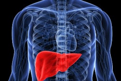
Hepatic steatosis is a common condition, and can lead to cirrhosis in 20% of patients, Schwartz told session attendees. MRI is typically used as the reference standard for diagnosing hepatic steatosis, but since CT is a common imaging exam, it offers an option for opportunistic screening, she said.
Schwartz's group sought to compare the performance of photon-counting CT with MRI for liver fat quantification in a simulated population of obese patients. The study used phantoms with inserts that consisted of six different concentrations of fat (from 0% to 100%) and fat-iodine mixtures (0% to 50% fat plus 3 mg/mL iodine); the group imaged the phantoms with a photon-counting CT device and a 1.5-tesla MRI system. But it also included 12 obese patients (mean body mass index, 30.3 kg/m2) with fatty liver disease, all of whom underwent an MRI and a photon-counting CT of the abdomen on the same day.
The study found that photon-counting CT showed comparable performance to MRI for identifying fat percentages, both on the phantom research side and on the patient side. The team found no significant differences between the known fractions of fat (p = 0.32) or iodine (p = 0.6) in the MRI-measured concentrations compared to the PCCT ones of the phantoms. This trend appeared among the patient data as well, with a mean signal fat fraction on MRI of 14.5% and on photon-counting CT of 11.8% -- a non-significant difference (p = 0.138).
Schwartz did concede that the research is quite preliminary, as it included only a small patient sample and the team did not test the use of intravenous contrast. But the results are promising.
"Our preliminary analysis of the performance of photon-counting CT compared to MRI for quantifying liver fat shows good correlation between the modalities," she said. "And the fact that in the phantoms the fat fraction analysis was very precise when iodine was also present bodes well for imaging patients with intravenous CT contrast."