PET/MRI has identified which compartments of the knee may be the best targets for new treatments for early osteoarthritis, according to a study published May 16 in Clinical Radiology.
Indian researchers in New Delhi found uptake of the radiotracer F-18 sodium fluoride (F-18 NaF) on PET/MRI scans by bone marrow lesions and osteophytes -- two frequent imaging findings in osteoarthritis -- varied by knee compartment. The study results could help researchers design new treatments that target early bone degeneration associated with the disease, the authors wrote.
"Using PET to measure bone metabolic activity alterations in conjunction with morphological changes from MRI could provide new insight into [osteoarthritis] assessment," wrote first author Dr. Amarnath Jena, a nuclear medicine physician at Indraprastha Apollo Hospitals.
Osteoarthritis is among the most common degenerative bone diseases in the world and causes pain and stiffness, especially in the hip and knee joints. Studies have shown that the presence of bone marrow lesions on MRI can increase the risk of cartilage loss, while osteophytes are associated with loss of joint space and are commonly used to diagnose the disease.
Both bone marrow lesions and the development osteophytes are generated by bone remodeling, a process triggered by damaged joints to cope with loading stresses.
PET/MRI has recently emerged as a new technique for exploring the complex pathophysiology of osteoarthritis, based on its ability to provide both bone metabolic activity alterations and morphological changes.
In this study, the researchers aimed to show that PET/MRI can identify compartmental differences between bone marrow lesions and osteophytes in the knees of osteoarthritis patients based on metabolic uptake values of F-18 Sodium Fluoride (NaF), a known marker of bone remodeling.
The group recruited 16 patients (32 knees) from January 2021 to July 2021 with no prior history Prior to the study, x-rays were acquired and patients were divided into three groups based on standard Kellgren-Lawrence (KL) osteoarthritis severity grading: patients without pain with KL grade 0 (13 knees), patients without pain with KL score > 0 (six knees), and those with pain with KL grade > 0 (11 knees).
Images of both knees were acquired on a PET/MRI system (Biograph mMR, Siemens Healthineers) and analyzed by a musculoskeletal radiologist and a nuclear medicine physician, both with more than 15 years of experience. Bone marrow lesions and osteophytes were scored using MRI Osteoarthritis Knee Score (MOAKS) criteria and corresponding maximum standardized uptake (SUVmax) values.
In the 32 knee joints analyzed, 53 bone marrow lesions and 177 osteophytes were identified. Bone marrow lesions and osteophytes both showed statistically significant differences among knee compartments, with SUVmax for bone marrow lesions and osteophytes highest in the medial tibia (MT).
In addition, the mean SUVmax increased with the grade of bone marrow lesions, from 2.7 for grade 1 lesions up to 48.5 for grade 3 lesions. SUVmax was highest in lesions in the MT, followed by those identified in the patella, medial femoral condyle, trochlea (Troc), lateral patellar facet-medial patellofemoral ligament (LPF-MPF), and the lateral collateral ligament (LT).
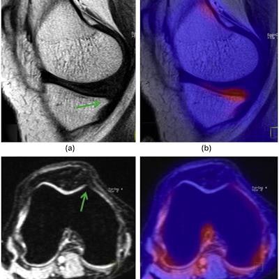
 A 30-year-old woman with bilateral knee pain and grade 1 osteophytes in both MT (a) sagittal T1 TSE fat-suppressed and Troc (c) reformatted axial 3D MEDIC compartments, but corresponding fused PET/MRI images (b,d), showed that MT osteophytes had higher SUVmax value (2.54) than the Troc osteophytes SUVmax value (0.73). Image courtesy of Clinical Radiology.
Similar to the bone marrow lesions, a proportional increase in SUVmax was also observed depending on the size of osteophytes, ranging from 1.3 for grade 1 osteophytes up to 6.64 for grade 3 osteophytes. SUVmax was highest in osteophytes in the MT, followed by those in the LPF, MPF, patella, LT, and Troc.
"F-18 NaF-PET/MRI showed that [bone marrow lesions] and osteophytes had different uptake values due to bone remodeling among the various knee compartments," the researchers stated.
This study was the first to attempt to determine the compartmentalized difference among bone marrow lesions and osteophyte uptake values on PET/MRI imaging, and has certain limitations, including a small sample size, the authors noted.
"The regional variation in the rate of bone remodeling should be explored in a larger cohort so that the factors responsible for this observation can be uncovered to help design disease-modifying osteoarthritis drugs in the future," Jena and colleagues concluded.