Interstitial Lung Disease in Rheumatoid Arthritis
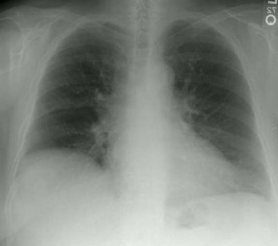
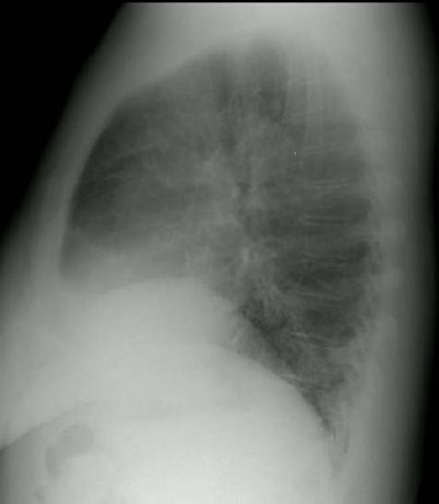
The frontal and lateral views of the chest are from a patient with rheumatoid arthritis. The elevation of the right hemidiaphragm is a chronic finding in this patient. The films demonstrate a shaggy appearance to the heart border on the frontal examination and prominent interstitial markings are very evident over the spine on the lateral examination. See CT scan below.


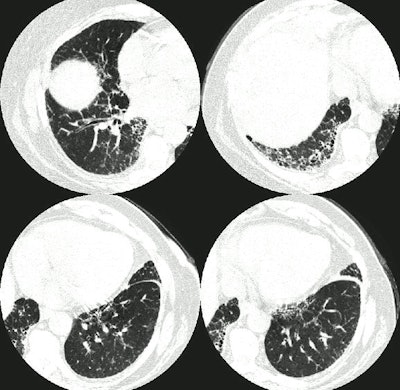
The CT scan demonstrates interlobular septal thickening and the presence of "honeycombing" within the lung bases- most pronounced in the right lower lung.
