Pulmonary Anatomy and Physiology:
There are approximately 350 million alveoli. Each one of them is enmeshed by a capillary web, arising from precapillary arterioles (10-30 microns in diameter). The blood flow through the capillaries depends on the difference of capillary pressure and a compressive effect of atmospheric pressure in the alveolus.
In the upright position, the capillary pressure above the level of the pulmonary valve is very low (with negative pressures in diastole), hence, atmospheric pressure compresses the capillaries and obstructs the blood flow. At the very most, blood flows briefly in that lung zone during peak systole. The capillary pressure rises towards the base under the influence of hydrostatic pressure of the blood column, progressively overriding the compressive effect of atmospheric pressure in the alveoli. Hence, the blood flow increases from apex to base quite rapidly. The ventilation also increases from apex to base (deeper diaphragmatic excursion produces a larger change in alveolar volume in the bases), but at a much slower rate as compared to the changes in blood flow. The result is a dropping ventilation perfusion ratio (V/Q) from apex (V/Q = 3/1) to base (V/Q = 1/1.6). In the recumbent position this hydrostatic gradient decreases and is reoriented to an antero-posterior plane. Increased perfusion to the upper lobes in an upright patient is suggestive of CHF, increased left atrial pressure, or alpha-1-antitrypsin deficiency.
Collateral ventilation in the lungs is supported by the pores of Kohn (communications between alveoli) and the canals of Lambert (communications between the respiratory bronchiole and alveolar ducts). This permits ventilation of the peripheral lung and often prevents collapse of areas distal to airway obstruction. However, lack of ventilation in those areas, given preserved perfusion, results in a functional (or physiologic) pulmonary shunt. It can cause severe hypoxia which is resistant to even 100% Oxygen inhalation, because administered oxygen cannot reach the capillaries at the effected site. However, very low alveolar pO2 values stimulate alveolar receptors and cause compensatory hypoxic vasoconstriction, redirecting the blood flow from poorly aerated lung to regions with better gaseous exchange. Thus, an area of decreased pulmonary perfusion is not specific for pulmonary embolism. Any parenchymal disease process such as pneumonia, alveolar edema or hemorrhage, pulmonary infarction, or tumor will produce a perfusion abnormality, but these conditions typically have an associated ventilation abnormality which produces "matching defects."
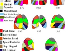
It is critical to keep a vivid image of pulmonary vascular segmental anatomy in mind for a proper interpretation of V/Q lung scans. Most of these charts are based on bronchopulmonary anatomy, recognizing a common derivation of left apical and posterior bronchi from the apico-posterior division of the upper lobe bronchus. Hence, there is a single apico-posterior bronchopulmonary segment on the left which is mirrored by two segments (apical and posterior) on the right. Vascular segmentation is identical on both sides (separate segmental artery feeding apical and posterior segments). Our chart is based on vascular segmentation, which is simplified for practical purposes.

