
Editor's note: As part of the celebration of AuntMinnie.com's upcoming 25th anniversary, we're presenting 25 for 25 -- a series featuring our most popular content for each of the last 25 years. New articles will be published each Monday until our official anniversary at RSNA 2024. In 2006, our top article shared the story of a radiologist whose life was saved by a 64-slice CT scanner just installed at the hospital he worked at.
Buying a major piece of imaging equipment is always a big moment for a radiologist. So Benjamin Roach, MD, was truly excited when his facility, Our Lady of Bellefonte Hospital in Ashland, KY, decided to purchase a new CT scanner. For the purchase of a cutting-edge, state-of-the-art 64-slice scanner, the excitement was palpable: "Think of the lives we'll save," he thought. Little did he know that one of the first lives saved would be his own.
Almost a year separated our purchase of a new 64-slice CT scanner (LightSpeed VCT, GE Healthcare, Chalfont St. Giles, U.K.) from the day we'd actually get to use it. Everyone knew the potential of this new technology, and we wanted to make sure we were in a position to capitalize on the new clinical functionality as soon as the scanner was ready.
Its speed would allow us for the first time ever to acquire vivid images of the heart, and, through postprocedure processing, generate additional views of the coronary arteries that supply the heart. This new CT would give us another tool in our technology arsenal for fighting cardiovascular disease.
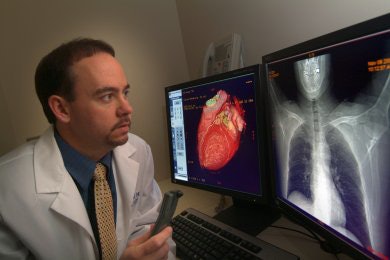 |
| Benjamin Roach, MD, of Our Lady of Bellefonte Hospital in Ashland, KY. Image courtesy of Benjamin Roach, MD. |
In anticipation, the entire radiology department embarked on a rigorous training program. We attended training sessions, conferences, and lectures. I was fortunate enough to participate in a visiting fellowship with David Dowe, MD, whom I consider the father of cardiovascular CT.
We began training on our newly installed VCT by scanning patients (employees and family members) who had one or more risk factors for heart disease: high blood pressure, high cholesterol, diabetes, smoking, and a family history of heart disease. Of all these risk factors, family history may be the most significant. All the others can be mitigated through behavior modification (eating right, exercising) or medication. I knew my family history and was worried about my brother.
Our family has a history of heart disease on both my mother's and father's side. My father underwent bypass surgery and has stents. But, I was concerned about my brother. Besides a sedentary lifestyle, he has two of the other risk factors: high blood pressure and high cholesterol.
I wasn't as concerned about myself. I'm 38 years old and take good care of myself. I've always been a runner. I had slacked off over the last few years because of time pressures at work but over the last year or so I've been running 4 to 5 miles pretty much every day. My blood pressure and cholesterol have always been borderline high, but diet and exercise have been keeping them under control. My father was pressuring me to scan my brother, and so we did.
We found that he had the early changes of heart disease. He was beginning to lay down soft plaque. But there were no areas of real stenosis, and certainly nothing that warranted intervention. I was so excited to be able to find this for my brother -- he could now choose to control this disease before it was too late.
Early detection is key
In any disease, the key to winning the fight is early detection. For example, breast cancer deaths have been on the decline partially due to better treatments but more importantly because of earlier detection. We knew the 64-slice scanner could detect not only significant disease but also disease in earlier stages before it progressed. This study provided my brother with knowledge about his disease. He now could make choices about his lifestyle and treatment to prevent further progression.
Most people at this early stage don't get choices when it comes to heart disease. More than 50% of people with heart disease do not initially present with the classic symptoms (chest pain, left arm pain, shortness of breath). In fact, most people with heart disease present with advanced disease causing either sudden death or a heart attack -- long past the point when they could have prevented it.
A heart attack permanently damages the heart. While the majority of advances in heart disease have focused on treatment, with technologies such as angioplasty, stents, and bypass procedures, 64-slice CT is the first major advance in the area of noninvasive diagnosis for heart disease. We wanted to not only detect the advanced disease but also find the early disease and treat it before it had the chance to advance.
Passing the stress test
My brother was OK, what about me? I had undergone a treadmill stress test a year or so ago. I had some infrequent twinges in my chest when I was stressed. I never had the twinges when running. In fact, I felt great during and after running. I took the stress test anyway. Being a runner, I blew the test away. The stress test concluded that I had no heart disease.
Nevertheless, I wanted to scan myself. I knew I had the one major risk factor (family history) but after seeing my brother's results, I wasn't that worried. I figured that -- worst case scenario -- I'd be at the same stage as he was and I'd go see my doctor and discuss medication to better control my cholesterol.
It was a Thursday when I was scanned. My partners, Dr. Eugene DeGiorgio and Dr. Steven Woolley, supervised the exam. As the initial images came up, they thought that there was probably some plaque present. I got off the table and waited with them for a couple of minutes until the detailed vascular images were reconstructed.
When the images came up we immediately saw one area of minor interest in my left coronary artery, and two areas of more significant interest in my right coronary artery, one mid and one distal. Occasionally, artifacts can be introduced into the images as a result of patient motion or through the reconstruction software. There are techniques to clear the artifacts, and so we began to attempt to clear them out of the images.
After about 10 minutes of work, the two areas remained. I just couldn't believe that I had what looked like two significant blockages of at least 70%. I asked Dr. Woolley to let me process the study. After additional work, Dr. Woolley said, "If this was any other 38-year-old patient, you'd be done with the exam and be recommending catheterization without hesitation." I realized he was right.
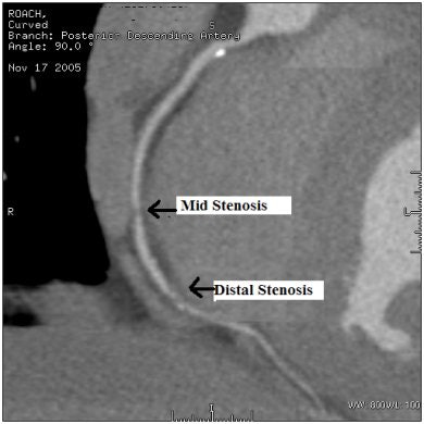 |
| 64-slice CT angiography image demonstrates 70% stenosis in Dr. Roach's mid and distal right coronary artery. Image courtesy of Dr. Benjamin Roach. |
Fortunately, as Dr. Woolley left the radiology department he ran into one of the hospital's interventional cardiologists. After relaying the findings of my exam, the cardiologist mentioned that they could perform a coronary catheterization at 7:30 a.m. the next day, which was Friday. I put all the images on a disk and finished the preop lab work that same day. I originally had plans on Friday to go furniture shopping with my wife, but suddenly my priorities shifted from a new couch to a needy heart.
During my 12 years as a radiologist, I received training in interventional procedures. I was fully aware of what was going to happen to me as I lay on the table that Friday morning. I was awake during the procedure and watched the images of my left coronary artery.
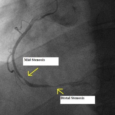 |
| Cardiac cath image demonstrates correlation with CTA. Image courtesy of Dr. Benjamin Roach. |
The interventional procedure confirmed the CT findings: there was minor plaque buildup in my left artery, but not enough to warrant a stent. Then the cardiologist began imaging my right artery. I saw him pass the areas of interest once, then double back to look again. Again the CT findings were confirmed. I had two significant blockages of almost 90%. Ten minutes later they were both stented and wide open. Three days later I was back to 100% and watching my son's football game.
While the new 64-slice CT scanners can find changes in heart disease, one of the best things about it is that its negative findings are 99% predictive. That is, if no plaque is found then you can be 99% certain that there is no plaque. That is extremely accurate and gives patients a tremendous sense of relief.
If the findings should come back positive, then the patient and his or her physician have knowledge and the power of choice. They can treat the disease with lifestyle changes and medication, or the findings may warrant a more aggressive approach such as catheterization.
Now I run 4 to 5 miles every day without fail. I never miss a day of running. Not only did the CT scanner impact my family, it impacted me. It saved my life.
Dr. Roach and Our Lady of Bellefonte Hospital would like to thank imaging services consulting firm Ivy Ventures of Richmond, VA, for its assistance in purchasing its 64-slice CT scanner. For more information about Ivy Ventures visit www.ivyventures.com.
The comments and observations expressed are those of the author and do not necessarily reflect the opinions of AuntMinnie.com.
Copyright © 2006 Ivy Ventures, LLC
