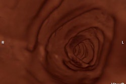
One of the latest trends in gastrointestinal imaging is like a scene from the 1966 sci-fi movie Fantastic Voyage: Patients swallow a small capsule outfitted with a miniature camera that snaps pictures as it passes through the small bowel. From its origins in Israel, capsule endoscopy has made the passage to India, where researchers at a Hyderabad hospital are putting it through its paces.
Capsule endoscopy is based on research conducted by Israeli rocket scientist Gavriel Iddan and U.K. gastroenterologist Dr. Paul Swain. The technology has been commercialized by Given Imaging Systems of Yoqneam, Israel, as the M2A Diagnostic Imaging System.
The Indian angle to capsule endoscopy comes from the software used to analyze capsule endoscopy images, which was developed in Hyderabad, according to gastroenterologist Dr. Nageshwar Reddy. Reddy is director of the Asian Institute of Gastroenterology in Hyderabad.
Reddy reported in his group’s experiences with capsule endoscopy in a presentation at the IRIA conference. Their study was recently published in the January 2004 issue of Journal of Gastroenterology and Hepatology (Vol.19:1, pp. 63-67).
Given's M2A capsule measures 2.6 x 1 cm. In addition to a digital CMOS camera, the capsule includes a transmitter that sends images via radio-frequency link to a data recorder worn on the patient's hip, where images are stored until they can be downloaded in the physician's office. The capsule takes two images per second over the course of eight hours as it travels through the gastrointestinal tract, for a total of about 60,000 images. Patients pass the capsule naturally.
In their study, Reddy's group examined 24 patients (21 men, three women, mean age 44.9, range 15-73 years). All underwent capsule endoscopy after routine endoscopy failed to provide a diagnosis.
Gastrointestinal bleeding was the most common indication for capsule endoscopy referral, with nine patients reporting this condition and another three reporting anemia. For the patients with GI bleeding, other studies -- esophago-gastro-duodenoscopy, colonoscopy, enteroscopy, and barium series -- were all normal.
Of the nine patients with GI bleeds, capsule endoscopy successfully demonstrated the abnormality in the small bowel in six studies. In three cases of these multiple angioectasiea, the accessible lesions were treated endoscopically using argon plasma coagulation during a subsequent push enteroscopy exam.
One patient with active bleeding was found to have a submucosal leiomyoma, which was operated on. Another patient with a submucosal lesion refused surgery and is being followed, without further complications.
The capsule did experience some complications in one GI bleed patient. This patient had a distal ileal stricture and a diverticulum. After 12 hours, the capsule remained stuck in the distal ileal region and the patient began to exhibit signs of small bowel obstruction.
He was operated on and the capsule removed, and at the site there was evidence of Meckel's diverticulum and an ileal stricture at the site with proximal ulcerations. The patient subsequently recovered well, Reddy said.
There were three normal studies, and in two of those, the patients went on without further bleeding episodes. In the third, the patient had a massive hematemesis two months later; a laparotomy revealed a 4-cm large extraluminal leiomyoma in relation to the proximal jejunum with small ulcerations on the mucosal surface.
In the three anemia patients, one patient had multiple hookworms. Another had an extra-hepatic portal venous obstruction in which esophago-gastric varices were earlier obliterated endoscopically, but the patient continued to experience anemia. The other patient had previously been operated on for pancreatic cancer.
The capsule was also used on a group of patients with unexplained weight loss and chronic diarrhea. The M2A capsule was able to detect multiple small intestinal ulcers with necrotic bases. Four patients had apththoid ulcers typical of Chron’s disease, while another patient had multiple deep linear ulcers in the jejunum and ileum.
As the study findings suggest, the primary indication for capsule endoscopy is obscure gastrointestinal bleeding, Reddy said. The group’s findings indicate that capsule endoscopy is twice as effective in enabling a diagnosis as push enteroscopy: 11/20 patients (55%) for capsule endoscopy versus 6/20 (30%) for push enteroscopy.
Chronic diarrhea is another possible indication. The capsule has been used for undiagnosed chronic abdominal pain, but has not performed as well in this indication, Reddy said.
One potential complication with capsule endoscopy is the possibility that the capsule might get stuck, such as what occurred in the diverticulum patient mentioned above. In cases like this, Given has a test capsule available that can be sent through the patient’s small bowel before the regular capsule. If the test capsule becomes stuck, it simply dissolves and is passed by the patient, Reddy said.
A major limitation of the capsule is the inability to control or steer the capsule as it moves through the small bowel. Other limitations include a slow passage time, large volume of images produced (and correspondingly long image review time), the likelihood that bowel contents could obscure images, and the lack of a therapeutic component. The capsule is also quite expensive (20,000 rupees, $450 U.S.), which could limit its use in India, Reddy said.
A new capsule that Given is working on could address some shortcomings, Reddy said. The capsule would both be steerable via remote control and could include a therapeutic component, such as the ability to attach clips or inject a scleroting drug.
By Brian Casey
By AuntMinnie.com staff writer
March 12, 2004
Related Reading
Capsule endoscopy fares well in small bowel versus CT, barium enema, January 14, 2004
Copyright © 2004 AuntMinnie.com


