India is not far behind the developed world when it comes to adopting new technology for patient care and treatment. This was proved yet again when Dr Jayaraj Govindaraj successfully completed the second radiofrequency (RF) ablation procedure for osteoid osteoma on Tuesday.
In July, Govindaraj, senior consultant in the department of radiodiagnosis and imaging at Apollo Speciality Hospital, Chennai, completed what was probably the first RF ablation procedure for bone tumour in India. And following its success, Govindaraj performed a second procedure on a 34-year-old patient on Tuesday.
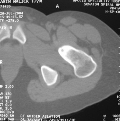 |
| Seventeen-year old patient with tumour in the neck of the femur. All images courtesy Dr Jayaraj Govindaraj, Apollo Speciality Hospital. |
Studies have shown RF ablation to be an effective technique for treating osteoid osteoma (Radiology, October 2003, Vol. 223:1, pp. 171-175). RF ablation is a minimally invasive interventional procedure performed under imaging guidance. It uses targeted radiofrequency energy to excite molecules, causing temperature to rise in the immediate vicinity of the RF probe and destroy tumour cells.
In the procedure Govindaraj performed, CT was used to localise the lesion, and axial slices of 2-mm thickness were obtained. Percutaneous RF ablation was performed under CT guidance, the RF probe was introduced into the nidus, and the tumour tissues were heated to a temperature of 100 degrees centigrade for three to five minutes.
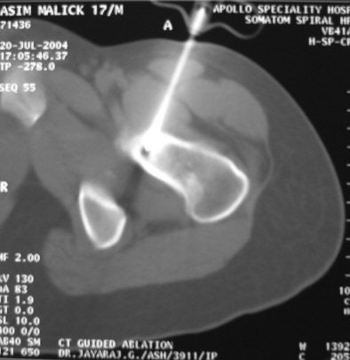 |
| RF ablation being performed under CT guidance. |
The unit used for the procedure was a Model 1500X RF generator and a Starburst SDE 16.5-gauge probe (RITA Medical Systems, Mountain View, CA).
The ablation was done two to three times at different angles for close to half an hour. The entire procedure takes about two hours. "The immediate effect is not apparent on CT, but when the patient recovers from anaesthesia, he is pain-free," Govindaraj told AuntMinnieIndia.com in an interview.
Osteoid osteoma can resolve on its own, but in some individuals it can persist, causing extreme pain. Surgery, which is an option in such cases, is done under imaging guidance using C-arm fluoroscopy. A larger amount of tissue has to be removed because the lesion cannot be localised as well as it can be done on CT, according to Govindaraj.
RF ablation offers an alternative that is accurate and less-invasive. However, caution should be exercised because bones conduct heat longitudinally and run the risk of becoming brittle when RF ablation is done. To minimise this risk, Govindaraj advises patients take complete rest for five days following the procedure, then avoid activities that put weight on the affected bone for about four weeks.
In instances when the tumour is in the lower limb, as is frequently the case, patients should walk with the support of crutches during the recovery period.
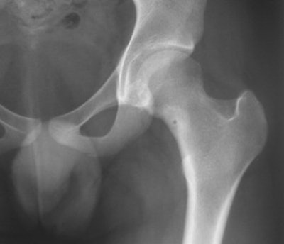
Recovery was complete for the 17-year-old patient on whom the procedure was first performed. "The patient had no pain, and the little foci had closed," Govindaraj reported. Earlier, when bone trephining was performed in January, the tumour had recurred.
 |
| Femur image three months after the procedure. |
Govindaraj will present the case as a poster at the upcoming 6th Asia-Pacific Congress of Cardiovascular and Interventional Radiology to be held in New Delhi from October 31 to November 3.
By N. Shivapriya
AuntMinnieIndia.com staff writer
October 28, 2004
Copyright © 2004 AuntMinnie.com

