How do you make sure a fluoroscope is warmed up? How about putting your hand in front of it until your finger bones appear? As crazy as that sounds, that's how early physicians approached the new x-rays discovered by Wilhelm Conrad Roentgen. Many of these doctors became radiology's first martyrs.
The excitement surrounding Roentgen's discovery of "a new kind of rays" in 1895 prompted hundreds of doctors to obtain the technology needed for making x-ray images of the patient anatomy. Their enthusiasm, however, was soon tempered by the growing awareness that x-ray could have serious health consequences if used improperly.
"Within 90 days of Roentgen's discovery, suspicion aroused in the minds of many investigators that x-rays or something involved in the production of x-rays might have some ill-effect on living tissues exposed to them," wrote Percy Brown, a leading early Boston radiologist, in his 1936 book American Martyrs to Science Through the Roentgen Rays. The book chronicled the American doctors, technicians, manufacturers, and nurses whose occupational exposures led to harmful injuries and deaths in the early years.
Even before Roentgen's discovery, he and other physicists had been exploring the insertion of electricity into glass vacuum tubes. The electric current colliding with the edge of the tube or any metal attachment produced invisible x-rays that ranged in all directions in any experimental laboratory. There was no concept of recognizing or measuring the invisible and unknown beams, even after Roentgen's discovery provoked the excitement of doctors in Europe and North America within weeks.
Indeed, it was not until 1928 that two committees were established at the second International Congress of Radiology to develop measurements of x-ray beams and exposures and to recommend techniques for protecting persons who were exposed to medical x-rays intentionally.
Starting in 1896, just weeks after Roentgen's first paper drew worldwide excitement, hundreds of doctors had obtained electric generators, glass vacuum tubes, and photo glass plates to examine patients for anatomy and make images of the x-ray exposures. By the spring of 1896, Thomas Edison, the American inventor of electric devices, had created a handheld fluoroscopic device, which many doctors welcomed and used regularly.
In those beginning weeks, some doctors noticed that patients with skin problems who were exposed to x-rays showed amelioration of their condition. But physicians also began to notice that their regular and frequent exposures to x-ray examinations were causing skin changes on their hands, rashes, loss of fingernails, and dermatitis problems.
The glass tubes were mounted with no form of protection for users and sprayed beams in all directions. When the fluoroscope was to be used, the tube was warmed and the doctor held his hand between the tube and fluoroscope.
When the doctor saw his finger bones, the device was considered to be ready, and the patient was then placed between the tube and the handheld fluoroscope. And sometimes an x-ray photo was exposed and the doctor had to develop the image on the glass plate. Some early x-ray users tried wearing silk gloves, coating their hands with salves and even searching out surgeons to remove affected tissues.
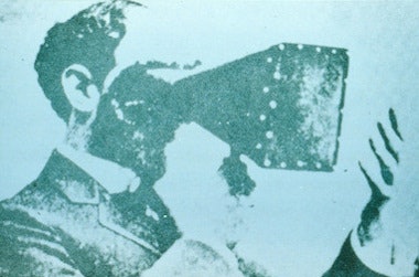 |
| Early fluoroscope operators made sure their machines were warmed up by checking to see if their finger bones appeared in the scope. Image is from American Martyrs to Science Through the Roentgen Rays, 1936. |
In the same early years, physicists and engineers in both North America and Europe experimented to improve the glass tubes to improve medical x-ray techniques. But some of them exposed themselves by demonstrating their devices to potential buyers. And some received the same exposures as the self-styled doctors using x-ray devices.
In Boston, one of the first physicians to start using x-rays was Dr. Francis Williams, a Harvard University internist at Boston City Hospital. His brother-in-law, William Rollins, took scientific interest in x-ray devices and came up with protection methods. Lead shields were known to block x-ray beams.
Rollins devised a wooden box, lined with layers of lead paint and with only one opening for the emission of an x-ray beam. He also advocated the use of lead shields and aprons for doctors to wear. But he did not promote his products aggressively, and a majority of early x-ray users never heard about them or took other precautions in the first two decades.
The first woman reported to die from performing medical x-ray exposures was Elizabeth Fleischman of San Francisco. Her physician brother-in-law, Dr. Michael J. F. Woolf, wanted to employ an x-ray device in his practice, and she became involved with its use. She gained a reputation by examining soldiers who returned San Francisco after being hurt in the Philippines in the Spanish-American War in 1898.
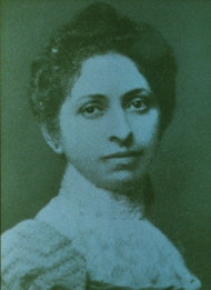 |
| Elizabeth Fleischman performed some of the first x-rays in California -- and was one of the first to die from overexposure to medical radiation. Image is from American Martyrs to Science Through the Roentgen Rays, 1936. |
"The fingers of both (her) hands were found to be badly ulcerated, chiefly the tissues over the middle phalanges and the middle joints ... the surfaces presenting both an ulcerated and warty condition, the warts assuming the form of necrogenia -- healing alternating with ulceration. All the secreting glands and hair-follicles were destroyed so that the skin was hard and dry and cracked easily. All forms of treatment by ointments and washes were without permanent benefit," wrote Brown.
His summaries of the experience and harm to all of the 27 x-ray pioneers profiled in his book were similar. The skin injuries led to chronic periods of vomiting, nausea, diarrhea, dermatitis, tuberculosis, and cancers in glands and organs.
Some of the pioneers faded as quickly as Elizabeth Fleischman. Dr. Walter Dodd, the developer of x-rays at Massachusetts General Hospital, lasted 20 years. Dr. Frederick Baetjer, the young surgeon who took up x-ray uses at Johns Hopkins Hospital, also had numerous surgeries and died in 30 years.
Some three years after his discovery and publication, Roentgen abandoned further x-ray studies. He experienced no ill health effects personally. Eighteen years later, the leading scientist at the General Electric Company laboratory, William Coolidge, devised a hot cathode ray device which made it safe for any x-ray users. The tube was housed in a metal container with one aperture for a focused beam of chosen patient anatomy.
The Coolidge tube in a decade replaced most of the unshielded glass tubes and handheld fluoroscopes. So radiology grew strongly, and its practitioners stopped suffering from x-ray exposures.
Otha W. Linton, MSJ, retired in 1997 as the associate executive director of the American College of Radiology (ACR) after 35 years. He also served as executive director of Radiology Centennial in 1995. Mr. Linton holds a bachelor's degree in journalism from the University of Missouri and a Master of Science in journalism from the University of Wisconsin. His work has been published widely in the U.S. and abroad, and he is a regular contributor to several journals including Academic Radiology, the American Journal of Roentgenology, Radiology, and the Journal of the American College of Radiology. He joined the ACR staff in 1961 and had a key role in its growth. Over the years, his responsibilities with the ACR included government affairs, public relations, marketing, publishing, industrial liaison, and international relations. Just before his ACR retirement, he became the executive director of the International Society of Radiology and continues in that role. Also, since his retirement, he has written and published 14 histories of radiology societies and academic centers.
Sources
Brown, Percy, American Martyrs to Science through the Roentgen Rays. 1936, Charles C. Thomas, Springfield, Illinois.
Gagliardi RA, McClennan BL. A History of the Radiological Sciences: Diagnosis. Reston, VA: Radiology Centennial Inc.; 1996.


