Understanding the most recent mechanisms of ballistic trauma and injury patterns can guide radiologists' interpretations, according to an electronic exhibit at the 2022 American Roentgen Ray Society (ARRS) meeting in New Orleans.
The exhibit highlighted key imaging findings and techniques and anatomic problems related to firearms and ballistic injuries. The first author on the exhibit was Jaykumar Nair of St. Michael's Hospital and University of Toronto in Ontario, Canada.
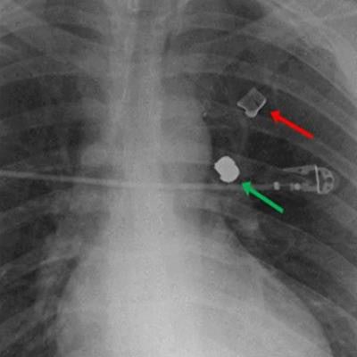
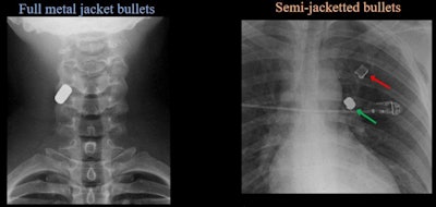
Several images illustrated the difference in impact between full metal jacket bullets versus semi-jacketed bullets.
 (Left) AP radiograph of cervical spine demonstrates single-density well-defined bullet within right posterolateral soft tissues of neck at C4 level, consistent with full metal jacket bullet (Right) Two different densities of ballistic material. Lower-density fragment in left upper lobe is copper jacket and high-density metal fragment is lead core, consistent with semi-jacketed bullet. Images courtesy of the ARRS.
"When interpreting imaging, one should use a mechanistic approach to establish bullet trajectory followed by assessment for injuries along the established path. Retained bullet fragments should be considered MRI conditional, and with the appropriate preceding workup patients, may be safely imaged,"


