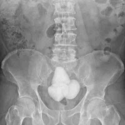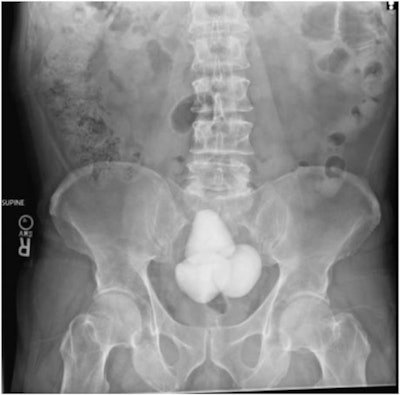
The authors of a case they dubbed "the Andes of bladder stones" described finding gigantic phosphate stones on a 60-year-old man's abdominal x-ray in a study published in Urology Case Reports.
The stones accumulated at an accelerated rate due to a rare case of the cumulative effects of both primary hyperparathyroidism (PHPT) and bladder outlet obstruction -- two distinct conditions that can cause urinary tract stone disease, explained doctors at the Dayton VA Medical Center in Ohio.
"No other similar cases have been described in the literature," wrote urologist Dr. Jonathan Hakim and colleagues.
According to the report, the patient had known bladder obstruction due to enlarged prostate and presented with difficulty performing intermittent catheterization due to pain. A subsequent kidney, ureter, and bladder x-ray revealed 13 centimeters of conglomerated radiopaque bladder stones. On review of imaging, the "impressive bladder stones" were conspicuously absent on x-ray 32 months prior, the authors noted.
 A KUB (kidney, ureter, and bladder) x-ray showing gigantic bladder calculi in a 60-year-old patient with known chronic urinary retention. Image and caption courtesy of Urology Case Reports through CC BY 4.0.
In addition, a nuclear medicine sestamibi parathyroid scan demonstrated increased radiotracer uptake within the right inferior parathyroid gland, confirming a diagnosis of primary hyperparathyroidism, the group noted.
The patient underwent prostatectomy, during which 254 grams of calcium phosphate bladder stones were removed. This was followed also by the removal of the offending benign parathyroid adenoma.
"On follow-up, the patient was voiding well with complete removal of bladder stones," the group wrote.
Ultimately, rapid accumulation of bladder stones over a 30-month period in the setting of both bladder outlet obstruction and PHPT is rare, the authors wrote. Nonetheless, the case highlights and emphasizes the role of a multidisciplinary approach to tackling these complex problems, they concluded.


