Who made it to the final round in this year's edition of the Minnies, AuntMinnie.com's event recognizing excellence in radiology? See below to find out who our expert panelists selected as the final candidates in this year's event.
The Minnies finalists were drawn from hundreds of candidates across 15 categories. You can also view a full list of candidates based on nominations from our members.
In the next round of voting, our expert panel will vote on the finalists, with winners announced in late October.
Most Influential Radiology Researcher
Emily Conant, MD, University of Pennsylvania Emily Conant, MD.
Emily Conant, MD, is a leading voice in breast imaging, lending her expertise to emerging technologies in breast cancer screening. Conant, a professor of radiology at the Hospital of the University of Pennsylvania (Penn), has led research examining the potential and role of digital breast tomosynthesis (DBT) and AI in breast cancer screening and diagnosis.
Conant has been named one of America's Top Doctors, recognized by Best Doctors in America, and also recognized in Philadelphia magazine's annual Top Docs issues. However, she is still seeking her first Minnies award. Could this be the year she finally achieves this accomplishment?
Conant earned her Bachelor of Arts in zoology from the University Of Vermont in 1976, but it was at Penn that her medical career started. She attained her Doctor of Medicine degree from the university in 1980 and has worn many hats for Penn since then.
Her current and previous roles at the university include being vice chair of faculty development in Penn’s radiology department, being a member of the Women's Health Scholar Certificate Program Advisory Council at Penn Medicine, and serving on several health and professional committees.
In March, Conant and colleagues had research published showing that DBT shows improved screening mammography outcomes over those seen in digital mammography in a study of over 1 million women. They found that DBT had lower recall rates and higher cancer detection rates in women compared with digital mammography.
Conant told AuntMinnie.com that these results suggest that DBT should be the standard of care for mammographic screening.
She also co-authored a paper published in June in MDPI Cancers that examined a clinical risk model for personalized breast cancer screening and prevention. The researchers found in their study of over 8,100 women that the model showed higher performance for both long-term and short-term risk assessment compared with imaging-only and Tyrer-Cuzick models.
Perry Pickhardt, MD, University of Wisconsin - Madison
Perry Pickhardt, MD, is no stranger to the Minnies, having won in this category in 2016. However, Pickhardt has been busy since then advancing research related to abdominal imaging.
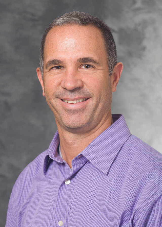 Perry Pickhardt, MD.
Pickhardt is chief of gastrointestinal imaging at the University of Wisconsin (UW) in Madison, as well as medical director of oncological imaging at the UW Carbone Cancer Center, a position he accepted in 2017.
He graduated from UW in 1991 with a Bachelor of Science in physics. Afterward, he graduated from the University of Michigan with his Doctor of Medicine in 1995 and then became a resident in diagnostic radiology at the Mallinckrodt Institute of Radiology at Washington University in St. Louis. It was here that he co-edited a textbook on body CT and published a number of scientific papers. Pickhardt later served in the U.S. Navy and organized a large multicenter screening trial evaluating CT colonography at the National Naval Medical Center in Maryland.
In February, Pickhardt co-authored research published in the American Journal of Roentgenology that assessed the technical adequacy of fully automated AI body composition tools in external CT exams. The researchers found that all three tools had high technical adequacy rates and wrote that their results support their generalizability and potential for broad use.
Pickhardt in 2022 spoke at the International Society for Computed Tomography (ISCT) annual meeting, speaking on the potential for opportunistic CT screening. He said that using additional clinical data on CT imaging could save healthcare costs, especially when AI tools are used as well. He cited research that found that AI-assisted CT-based opportunistic screening was a cost-effective strategy.
Pickhardt has received numerous accolades in his career. Aside from the 2016 Minnies award, he previously received the best paper award at the annual meeting for the Society of Gastrointestinal Radiology on four occasions. His work on abdominal imaging has led to over 400 scientific publications and book chapters, as well as multiple textbooks.
Most Effective Radiology Educator
Christine "Cooky" Menias, MD, Mayo Clinic Arizona
 Christine "Cooky" Menias, MD.
For the third year in a row, Christine "Cooky" Menias, MD, of Mayo Clinic Arizona has been nominated as a finalist for Most Effective Radiology Educator. A specialist in abdominal imaging, body CT and MRI, and emergency radiology, she brings decades of experience to her educational efforts as professor of radiology at Mayo Clinic in Arizona. In 2021, her educational activities further expanded when she began her tenure as editor of the RSNA's RadioGraphics journal.
Menias earned her medical degree in 1995 from George Washington University School of Medicine and Health Services in Washington, DC. She has served as professor at the Mayo Clinic for more than a decade; before arriving at Mayo, she taught at Washington University School of Medicine in St. Louis for 15 years.
She has often been recognized for her educational leadership. This year, Menias was honored with the Society of Abdominal Radiology's Gold Medal Winner award. Last year, the RSNA bestowed on her a Lifetime Honored Educator award and in 2021, she was named Educator of the Year for Research by the Mayo Clinic Arizona Resident and Fellows' Association. In 2019, she received the Distinguished Educator award from the American Roentgen Ray Society and a Distinguished Educator award from the American Roentgen Ray Society.
Katja Pinker-Domenig, MD, PhD, Memorial Sloan Kettering Cancer Center
 Katja Pinker-Domenig, MD, PhD.
Katja Pinker-Domenig, MD, PhD, is senior faculty in the department of radiology at Memorial Sloan Kettering Cancer Center and Memorial Hospital for Cancer and Allied Diseases, as well as professor of radiology at Weill Medical College of Cornell University, all in New York. She is director of research and breast MRI at Memorial Sloan-Kettering Cancer Centers' breast imaging service.
Her research interests include advanced breast imaging with high-resolution MRI using multiple advanced MRI parameters, hybrid imaging (PET/MRI) with specific tracers, and the application of AI in oncologic imaging.
Pinker-Domenig earned both her medical degree and her doctorate in Vienna, her hometown, at the Medical University of Vienna in 2003 and 2015, respectively. After serving a number of years as professor of radiology at that university she began work at Memorial Sloan-Kettering in 2014.
She serves on several editorial boards, including her current position as editor in chief of the British Journal of Radiology Open and deputy editor of the Journal of Magnetic Resonance Imaging.
One clinical accomplishment of which she is most proud is the successful implementation of a multiparametric breast MRI framework in routine patient care. After her arrival at Memorial Sloan-Kettering, she has continued to refine the use of multiparametric high spatial and temporal resolution functional breast imaging in clinical care, "implementing novel sequences for most accurate breast cancer detection," she said.
Most Effective Radiologic Sciences Educator
William Faulkner and Associates, Chattanooga, TN William Faulkner, RT.
It's not the first time William Faulkner, RT, has been acknowledged by a Minnies award: In 2002, he was voted "Most Effective Radiologic Technologist Educator." In 1998, he established his consultation firm, William Faulkner & Associates, through which he offers onsite MRI safety audit and risk assessment consultation as well as educational material regarding cardiac and neurostimulation devices that can be used with MRI.
Faulkner earned his Associate of Science in Radiologic Technology degree in 1975 from Chattanooga State Technical Community College and his RT certification that same year from the American Registry of Radiologic Technologists (ARRT). He went on to secure an advanced certification in MRI in 1995 and in CT in 1997. He is a credentialed MR safety officer and current chair of the American Board of Magnetic Resonance Safety (ABMRS); he was a founding member of this board in 2015 and first president of the Society of Magnetic Resonance Technologists (SMRT) in 1991.
In addition, Faulkner has served as editor of the International Journal of Neurology and has written extensively on topics ranging from gadolinium-based MRI contrast agents, the principles of radiographic imaging, and adjusting MRI scan parameters for patients with active implantable devices; a second edition of his book, Rad Tech's Guide to MRI: Basic Physics, Instrumentation and Quality Control, was published in 2020.
Joy Renner, RT, University of North Carolina at Chapel Hill
 Joy Renner, RT.Joy Renner, RT, is also a Minnies award returnee: She was the winner for this same category in 2006. Renner has made her career at the University of North Carolina (UNC) at Chapel Hill, earning a Bachelor of Science in Radiologic Science and a Master of Arts in Curriculum and Instruction there. Currently, she directs the university's division of radiologic science.
As an educator, she teaches courses ranging from "Integrated Principles of Radiographic Analysis" and "Clinical Decisions in Radiology" to "Issues in the Radiology Practice Environment" and "Musculoskeletal Imaging and Procedures." What she enjoys most about working with students is "helping them discover academic and career paths suited to their talents and interests where they had not considered before."
"My work at UNC as an educator and division director has allowed me to bridge the healthcare corporate world and education through workforce projects at all levels of education – from certificate and baccalaureate degree programs to master's programs," she told AuntMinnie.com. "I have seen a lot of change in healthcare needs and workforce trends over my four-decade career, and a key piece of staying viable is finding innovative ways to fund programs and to offer students enticing educational opportunities."
Most Effective Radiology Administrator/Manager
Andrew Menard, JD, Johns Hopkins Health System, Baltimore
 Andrew Menard, JD.
For the second year in a row, Andrew Menard, JD, has been voted in as a finalist for the Minnies' category of Most Effective Radiology Administrator/Manager, and it's easy to understand why. As executive director of radiology strategy and innovation for Johns Hopkins Health System in Baltimore, Menard tracks economic, political, and business trends that could affect both professional and technical radiology -- and then develops strategies to grow, support, and sustain the university's clinical care, education, and research missions. He previously served as Hopkins’ chief administrative officer for radiology.
Menard earned his law degree at Harvard and came to Hopkins in 2018 from Brigham and Women's Hospital, where he served as executive director of radiology. While at Brigham he helped establish a consortium to evaluate the impact of radiology clinical decision support on imaging utilization as part of the Medicare Imaging Demonstration, later providing input to draft legislation that eventually became the Protecting Access to Medicare Act (PAMA). He also assisted several academic radiology groups with their applications to become qualified provider-led entities under PAMA. He is also a member of the board of directors of the Harvard Medical School Library of Evidence. Additionally, Menard is a prolific writer, with his most recent paper, "Medicare Imaging AUC Program: Sometimes Less Is More" being published in August in Health Affairs.
Menard's efforts have been noticed: In addition to his nomination to the category this year and last, in 2015 he received the Association of Administrators in Academic Radiology (AAARAD) award for advancement of the profession of radiology.
Paulo Castaneda, Stanford Health Care, Stanford, CA
 Paulo Castaneda, RT.
Director of Imaging Services at Stanford Health Care Paulo Castaneda "loves tackling difficult problems and finding new opportunities for quality and process improvement," he said on his LinkedIn page. He holds an MBA from Western Governors University in Millcreek, UT, and earned his nuclear medicine technologist certification from the VA Palo Alto Technologist Training program.
Since he arrived at Stanford in 2007, Castaneda has served as a nuclear medicine/PET-CT technologist, a nuclear medicine supervisor, and a nuclear medicine manager. Before his tenure at Stanford, he worked as a researcher at the Ernest Gallo Clinic and Research Center in Emeryville, CA.
Castaneda helped facilitate the opening of an imaging clinic at Stanford Health Care's South Bay Cancer Center in 2015, and in 2021 helped open the university's Division of Nuclear Medicine and Molecular Imaging's Theragnostics clinic in Palo Alto, CA. He has received a number of awards during his career, including Stanford's Health Care Award for Special Contribution in 2017 and its Radiology Employee of the Year award in 2020.
Best Radiologist Training Program
Mallinckrodt Institute of Radiology, St. Louis, MO
The Mallinckrodt Institute of Radiology, part of Washington University in St. Louis, has been awarded the Minnie in this category four times, but it's been a decade since its last win.
Boasting one of the most prestigious and consistently top-ranked radiologist training programs in the world, Mallinckrodt offers hands-on mentoring and one-to-one instruction from leaders in radiology education, as well as plenty of opportunities for social networking. The institute also takes pride in its large network of radiology alumni, many of whom hold leadership positions in clinical care, research, and teaching.
The curriculum has trainees learn the ins and outs of different subspecialties in radiology, including neuroimaging, breast imaging, and pediatric imaging to name a few. They learn about best practices and uses for imaging modalities such as CT, ultrasound, and mammography. Along with that, they have opportunities to work in night float and on-call schedules, according to Mallinckrodt. The institute’s residency program also has three residency program tracks for interventional radiology, including integrated, independent, and early specialization.
During their final year of training, residents in the diagnostic radiology residency who are not doing an early specialization in interventional radiology, research, or diagnostic radiology/nuclear medicine track can complete one or more selective rotations, otherwise known as “mini-residencies.” Mallinckrodt also encourages residents who are involved in projects that require dedicated blocks of research time to pursue a research elective that provides uninterrupted time to work on mentored projects.
Finally, the institute promotes equity for all members through the university’s Office of Diversity, Equity, Inclusion and Justice. The office fosters a community that embraces and reflects the diversity of backgrounds, experiences, and perspectives of radiologist trainees, according to Mallinckrodt.
University of Pennsylvania, Philadelphia, PA
 A frequent finalist, the University of Pennsylvania (Penn) broke through in 2022 to win its first Minnie for Best Radiologist Training Program. Can it successfully defend its title?
The Ivy League school has multiple learning tracks and residency programs to go with its rich history in the medical field. The university launched the first medical school in the U.S. -- the College of Philadelphia, which was established in 1765 and is now the Perelman School of Medicine. It also had the first hospital in America (Pennsylvania Hospital), according to the institution.
Penn offers residency programs in interventional and diagnostic radiology, with the latter offering clinical and research tracks. It also offers aspiring imaging leaders specialty tracks in business and innovation, healthcare leadership, and informatics.
Taking advantage of modern teaching methods, the university also utilizes personalized teaching sessions, team breakouts, small-group sessions, dedicated minicourses, and flipped classrooms, among other methods, according to Penn. It also covers subspecialty areas, including physics.
Trainees can also utilize Penn Interventional Radiology’s multiple up-to-date interventional suites. These include one CT-fluoroscopy suite, two C-arm suites, two procedural CT scanners, a preprocedure suite, and a large postprocedure recovery suite. The Perelman Center, meanwhile, has two C-arm suites, a venous ablation suite, and a dedicated interventional radiology clinic with four clinic rooms and provider workroom areas. Ultrasound is also available in all interventional suites and clinics.
Meanwhile, residents work with a diverse case mix from multiple hospitals within and associated with Penn Medicine to gain clinical experience, the institution said. This includes reading radiographs, independent call shifts, and working in a night float system.


