In the 1966 science fiction film "Fantastic Voyage," a miniaturized Stephen Boyd nearly meets his death inside the left lung of a dying man, when the force of the patient's breathing hurls Boyd's microscopic body against an alveolar wall.
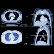
Happily, in the years since the movie was released, doctors have found more practical ways to explore the lungs and bronchi, with tools such as CT and bronchoscopy that have now become indispensable in diagnosing and treating pulmonary disease. In recent years, researchers have combined CT imaging with navigational concepts borrowed from bronchoscopy to create virtual bronchoscopy (VB), a noninvasive guided imaging technique that enables a sort of virtual fantastic voyage through the airways.
Researchers from the University of Iowa and Pennsylvania State University will discuss their joint research on VB at the upcoming Medical Imaging 2000 conference to take place February 12-17 in San Diego. Dr. Ronald Summers, a radiologist with the National Institutes of Health (NIH), will talk about his own team's research and clinical experience with VB. Brian Harvey, of the FDA's Center for Devices and Radiological Health, and Russell Kirchoff, of Olympus America, will round out the panel with regulatory and industry perspectives on the emerging technology.
The goal of the workshop is to better understand the state of art of VB, whether it is approaching the point where it can be introduced in routine clinical practice, and, if so, under what circumstances.
VB's origins
Since the first fiberoptic endoscopes became available in 1974, bronchoscopy has become a valued tool in diagnosing and treating major airway disease. The procedure, performed by inserting a thin tube through the patient's nose or mouth into the bronchi, allows the physician to traverse, see and photograph the inside of the bronchi to assess disease, and guide biopsy and surgery. Image capture improved with the introduction of digital CCD cameras in the mid-1990s.
But like all useful tools, bronchoscopy has its limitations. From the patient's perspective, the procedure can be lengthy and uncomfortable, making frequent follow-ups impractical. Complications, while rare, can include bacteremia and pneumonia, fever, localized bleeding, and even pneumothoraxis. Doctors must also deal with the physical limitations of the devices, which at 5-6 mm in width, can be used only in the widest airways. Inserting an endoscope through a narrowed bronchus would make breathing impossible, so the procedure cannot be used to gauge the length or extent of bronchial stenosis -- or see what lies beyond it.
A new approach
In contrast, nearly seven years of research point to VB's ability to transcend many of bronchoscopy's limitations. In independent research with the technique at the National Institutes of Health, Summers is impressed with VB's potential as a diagnostic tool.
"(Virtual bronchoscopy) is producing really incredible sections of airway structures and the effects of inflammation and tumors on them," Summers said. "And because it's noninvasive, it's amenable to frequent follow-ups. It's also useful for communicating with physicians and patients about the disease process, so you can show them what's going on, or how they can think about treating it, or how it's changing over time. The (CT scan) is done in two breathholds and it's very comfortable for the patient."
Summers said the most common airway disorders he sees in his patients involve bronchial inflammation and infection. "The diseases are kind of unusual: mycobacteria ... and a disease called Wegener's granulomatosis that causes inflammatory tissue to form around small blood vessels," Summers said.
Summers has previously published papers on visualization techniques using bronchoscopy, based on his clinical results at the NIH. He has evaluated about 70 patients to date, working with a team that includes a scientist, an engineer and a doctoral fellow. Moving beyond mere visualization of pathology, he said current research efforts are aimed at quantifying airway pathology for use in computer-aided diagnosis.
Quantifying the data
Quantitation of airway structure and pathology with VB is also the main focus of researchers at the University of Iowa, headed by Eric Hoffman, PhD, and Dr. Geoffrey McLennan, director of bronchoscopy at the university's department of internal medicine. McLennan has used the technique successfully on about 450 patients so far, in interventional as well as diagnostic applications.
Armed a with five-year NIH grant of about $2.5 million, the Iowa researchers will continue their long-term collaboration with computer scientists at Penn State, who are developing the software for the project. The main purpose of the NIH project, according to Hoffman, is to explore interventional applications for VB.
"Virtual bronchoscopy is part of a larger goal of developing tools for noninvasively assessing the lung," Hoffman said. "The main interest with the VB project was to link the real bronchoscopic exploration with the virtual bronchoscopy pictures. As the bronchoscopist moves down the airway, he can't see what's outside the airway, so by knowing where the tip of the bronchoscope is relative to the virtual bronchoscopic pictures, we can tell the bronchoscopist where to stop and poke across the airway to do (for example) a needle biopsy of a suspected lesion."
Precise measurements are critical for interventional applications of VB, Hoffman said, because the physician must know exactly where and how deeply to cut into a lesion, for example, while avoiding adjacent vascular structures. Measurement is also important in manufacturing the wire stents that are inserted in narrowed airways to hold them open. When stents are too small, they can be coughed up or sucked into the lungs, Hoffman said, while stents that are too large can fail to open fully, resulting in an overgrowth of tissue.
In order to turn CT image data into precise quantitative measurements, the researchers rely on software that is based on volumetric and dynamic imaging techniques first developed in the early 1980s. The heart of the program is an algorithm that determines the exact center of the airway. From this measurement, the software can calculate the dimensions of the airway at any point, the size and distance of adjacent structures, and can plot a precise path through the center of the airway.
Building the tools
William Higgins, PhD, is the computer scientist overseeing Penn State's multidimensional image processing laboratory, where software for the joint project with Iowa State is being developed. He said the lab's bronchoscopy software provides extensive quantitation that enables the user to know his exact location at any point in an airway. New for this project, he said, are multiple views that can be manipulated to provide the most clinically useful tour of the bronchi ahead.
"It shows sliced cuts in different directions," he said. "You can take the airway and open it up and straighten it out, or use a look-ahead view -- or construct a slim-slab view that gives you an idea of the tissues coming up, which is very important in trying to find lymph nodes or walls where cancer might be."
Higgins said the advanced quantitative abilities of the new volumetric software, combined with more powerful CT scanners, would ultimately render visual analysis of CT images obsolete. However, he said the payoff would be far better diagnosis of lung and bronchial disorders.
However, the University of Iowa's Hoffman cautioned that developing reliable quantitation software was only part of the picture; that widespread interventional use of VB would not be possible until more doctors are able to use the technology effectively.
"Physicians like Dr. McLennan can take those measurements and do something creative interventionally," Hoffman said. "Those sorts of physicians are few and far between."
For his part, Summers of the NIH said continuing improvements in CT scanners would help assure VB's place in clinical practice.
"In the coming years, CT scanners are going to give us better and better resolution, and we're going to pick up smaller and smaller airways, and I think that may lead to novel types of diagnoses," Summers said. "I see VB as an adjunct to looking at standard chest CT images -- and I think it's here to stay."
By Eric Barnes
AuntMinnie.com staff writer
February 11, 2000
Copyright © 2000 AuntMinnie.com


