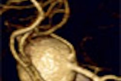
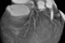
Early-enhancing hepatic pseudolesions can't normally be seen with ultrasound, but digital subtraction angiography (DSA) can usually find them, and CT does an excellent job of depicting their unique morphology.
Those are some of the conclusions of a 388-patient study presented the 2001 European Congress of Radiology in Vienna. Dr. Laura Romanini, from the Institute of Radiology at the University of Brescia in Italy, shared the results of her team's efforts to evaluate the frequency, morphologic features, and vascular patterns of hepatic pseudolesions.
Because pseudolesions caused by portal-venous impairments can be mistaken for true hepatic tumors, reliable methods of differentiating the two have been sought in a few smaller studies over the years, mostly in Japan.
"[Pseudolesions] are generally subcapsular or peripheral, mostly wedge-shaped, and all of the lesions had enhancement in the arterial phase, with a dot-light sign -- a small vessel -- in the lesion," Romanini said. "Generally these lesions are not recognizable in ultrasound studies. Most seem to occur as a result of cirrhotic changes in liver disease, the rest from transhepatic interventional procedures."
The so-called "dot light sign" signifies the presence of a markedly hyperdense vessel seen in the arterial phase, within the pseudolesion area of the liver parenchyma, Romanini said. The pseudolesions are typically not visible in the portal-venous enhancement phase.
For the study, 388 patients underwent spiral CT between March 1999 and August 2000. They were divided into three groups. Group 1 consisted of 248 patients with chronic hepatitis or cirrhosis; group 2 consisted of 59 patients with suspected benign focal liver lesions; and group 3 consisted of 81 patients with suspected hypervascular metastases.
Arterial-phase spiral CT images were acquired 25 seconds following bolus injection of contrast; portal-phase images were acquired 90 seconds following contrast administration.
Forty-seven pseudolesions were found in 27 patients, Romanini said. "Forty-two pseudolesions were found in the first group -- hepatopathic patients -- only two in the second (group), and three in the third."
Both pseudolesions in the second group of patients were found next to small capillary hemangiomas; both of these had a dot-light sign. Two of 3 pseudolesions in group 3 had the dot-light sign.
On CT, none of the pseudolesions were recognizable in the contrast phase, all of them were hyperdense in the arterial phase, 70% were hyperdense in the portal-venous phase, and 30% were slightly hyperdense in the portal-venous phase, Romanini said.
Seventy-eight percent of pseudolesions were isodense in the late phase, and the remaining 22 were slightly hyperdense. All of the pseudolesions were subcapsular, 80% were wedge-shaped, and 20% were round-shaped or oval-shaped. The characteristic dot-light sign was found in 74% of the pseudolesions.
No pseudolesions could be identified in the ultrasound exams. However, DSA performed in 14 patients with hepatocellular carcinoma showed 14 active portal shunts in the areas of pseudolesions imaged on CT. Three of the pseudolesions were biopsied, and CT follow-up in all patients within 18 months showed no changes.
The large number of pseudolesions found in the first group of hepatopathic patients seemed to confirm the group's hypothesis: that these pseudolesions are due to arterial-portal shunts caused by cirrhotic changes, she said.
Pseudolesions in groups 2 and 3 appeared to correlate with iatrogenic arterioportal shunts that occur during interventional transhepatic procedures, she said, "but we cannot exclude congenital arterioportal shunts, or nonportal shunts in the venous branch."
Other recent efforts to detect and differentiate pseudolesions are also worthy of note.
For example, a study in the Journal of Magnetic Resonance Imaging compared 3-D gradient-echo dynamic MR imaging (using 3-D FISP and double-dose gadolinium) with CT hepatic arteriography in 20 patients with hypervascular hepatocellular carcinomas (HCC). While the MR technique detected 98% of HCC lesions compared with CT as the gold standard, MR missed 57% of enhancing pseudolesions, according to author Dr. Kengo Yoshimitsu and colleagues (February 2001, Vol. 13:2, pp. 258-262).
A study published last year in Abdominal Imaging found that CT during arterial portography under temporary balloon occlusion of the hepatic artery (BOHA-CTAP) diminished 5 of 8 pseudolesions compared with conventional CTAP. The authors concluded that BOHA-CTAP "can reduce pseudolesions caused by portal venous impairments and enable the demarcation of true tumors" (November-December 2000, Vol.25:6, pp.583-586).
By Eric BarnesAuntMinnie.com staff writer
May 10, 2001
Click here to post your comments about this story. Please include the headline of the article in your message.
Copyright © 2001 AuntMinnie.com


