(Ultrasound Review) Both CT and ultrasound imaging are widely used in the diagnosis of appendicitis with a resultant lowering in the clinically negative appendectomy rate. In order to determine which technique was superior, radiologists at Hershey Medical Center in Pennsylvania conducted a comparative assessment of various CT techniques and graded compression ultrasound.
They studied 100 consecutive patients with suspected acute appendicitis, and the results were published in the American Journal of Roentgenology. Diagnostic results were compared with surgical findings when appendectomy was performed. When surgical intervention was unnecessary, a comparison was made with clinical follow-up over at least three months. They looked at inter- and intraobserver variability, patient tolerance, diagnostic confidence, technical accomplishment, and diagnostic performance.
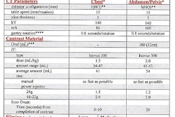
The diagnostic methods evaluated included "sonography, unenhanced focused appendiceal CT, complete abdominal CT using IV contrast, focused appendiceal CT with colonic contrast material, and repeated sonography with colonic contrast material," the authors stated.
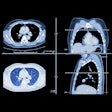
There were 24 patients (24%) with surgically proven acute appendicitis. Results demonstrated that sonography had a low sensitivity (34%) but high specificity (87%) and accuracy (74%). All of the CT methods outperformed sonography in sensitivity, specificity, and accuracy, and the reporting radiologists had more diagnostic confidence with CT. The appendix was more frequently demonstrated on CT regardless of the technique than during sonographic imaging.
The administration of colonic contrast afforded no advantage in diagnostic performance for either sonography or CT but improved CT confidence in positive cases. Patient tolerance was highest for unenhanced focused appendiceal CT and lowest for sonography and CT with colonic contrast, followed by graded compression ultrasound.
The study method could have contributed to the poor results for sonographic diagnosis. Because the ultrasound imaging was reviewed on video recordings, there was no communication between the sonographer and the interpreter, thus excluding important information regarding pain localization.
"Abdominopelvic CT gave the greatest confidence in cases with negative findings, and focused appendiceal CT with colonic contrast material gave the greatest confidence for cases with positive findings." The authors concluded that "a standard abdominopelvic CT scan is recommended as the initial examination for appendicitis in adult patients. However, focused appendiceal CT with colonic contrast material should be used as a problem-solving technique in difficult cases."
"Comparative assessment of CT and sonographic techniques for appendiceal imaging"Scott W Wise et al
Dept of Radiology, Pennsylvania State University College of Medicine, Milton S. Hershey Medical Center, 500 University Drive, Hershey, PA 17033, USA
AJR 2001; (April); 176:933–941
By Ultrasound Review
June 12, 2001
Click here to post your comments about this story. Please include the headline of the article in your message.
Copyright © 2001 AuntMinnie.com


