Discussions of CT calcium screening often focus on its technical aspects: image acquisition and reconstruction techniques, temporal resolution, ECG gating thresholds, and of course, the debatable merits of electron beam CT versus spiral CT.
But these matters, while important, are not everything. Still on the table are some very basic questions about the pathophysiology of atherosclerosis, and for that matter, the significance of calcium in the coronary arteries. How much calcified plaque is too much, and to what extent do its lesions foretell serious coronary events? The answers go to the heart of doctors' advice to patients, who undergo calcium screening to assess their odds of future problems. According to a leading cardiology researcher, the advice may soon change.
In a study presented at the 2001 RSNA meeting, Dr. Joseph Shemesh from the Sheba Medical Center in Tel-Hashomer, Israel, examined the different characteristics of coronary calcification in patients with acute versus chronic coronary events. The patients were a subgroup of the INSIGHT (International Nifedipine Gastrointestinal Transport System) calcification side arm study, which examined the safety and utility of calcium antagonists in patients with hypertension.
Shemesh and his colleague Dr. Michael Motro found that patients with the highest calcium scores had far fewer acute coronary events than those with less calcified plaque. A couple of previous studies had reached similar conclusions. And because acute events are the ones doctors most want to predict, the group's findings could potentially impact both the practice and the business of CT calcium screening.
"Coronary calcium is associated with a higher incidence of coronary events," Shemesh said. "However, differences of opinion exist as to whether or not calcification is associated with vulnerable and ruptured plaques, or is a sign of stable or unstable lesions, and whether it can predict the presence of plaque rupture. The extent and characteristics of coronary calcium -- the underlying lesions, acute versus chronic coronary events -- have not been clarified yet."
The study examined the extent and characteristics of calcific atherosclerotic lesions in acute versus chronic coronary events. Fifty hypertensive patients (mean age 66.6 years, 78% male) underwent ungated dual-slice spiral CT imaging using a Marconi Mx8000D dual-slice scanner (Philips Medical Systems North America, Bothell, WA).
All of the patients sustained a coronary event during the 3-year follow-up, and all had undergone spiral CT imaging within 12 months prior to the event. The coronary calcium (CC) score was calculated at the proximal 6 cm of the coronary tree.
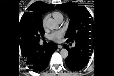 |
A total of 883 calcific lesions (defined as 0.5 mm or larger and > 90 HU) were identified in the patients. The researchers calculated the number of lesions, number of vessels with CC, total density of the lesions, and total CC score (TCS) for each patient. The presence of calcium was defined as TCS > 0, Shemesh said.
"As for the definition of acute versus chronic events...acute coronary syndrome manifests as unstable angina, acute myocardial infarction, and sudden death," Shemesh said. Acute events are dangerous and unpredictable, with underlying pathologies of plaque rupture and erosion, inflammation, thrombosis, and vasoconstriction.
"On the other hand, stable coronary disease, which we call chronic coronary artery disease, is mainly manifested with stable angina, which is flow restriction," he said. In these patients, coronary angiography often shows more than 50% lumen obstruction; percutaneous transluminal coronary angioplasty (PTCA) and coronary artery bypass grafting (CABG) are used to revascularize the vessels.
- The chronic events included acute myocardial infarction (n=20), unstable angina (n=6), acute ischemia (n=2), and the sudden death of one patient.
- The chronic (stable) patients had elective angiography (n=12), PTCA (n=2) CABG (n=2) and severe stable angina (n=5).
- According to the results, there was a high prevalence of coronary calcium (TCS > 0) in both the acute (93%) and chronic (96%) patient groups.
"However, if you look at the characteristics of calcium, you find a real big difference between these two groups," Shemesh said. "Coronary calcium in three vessels was three times more prevalent in the chronic group in the number of lesions, and total calcium was 15 times higher than in the acute group," he said. "The total area was 10 times higher in the chronic group than in the acute group."
 |
All of the patients who suffered acute events had calcium scores of less than 500. The median was 163.
"To summarize, a mild degree of calcification characterizes patients with acute coronary syndrome. However, larger, high-density calcified lesions are associated with chronic coronary events," he said.
While the primary goal of screening is to identify patients who are most likely to undergo acute coronary events, some of them may not be identified in screening, he said.
"If you stress these patients you will get negative stress tests, if you do angio on them you will get normal coronary arteries, or less than 50% stenosis," he said.
Moreover, the earlier, acute stage is where the atherosclerosis itself can be treated, for example with statin therapy. As for the chronic patients, revascularization simply treats the symptoms, not the disease itself, he said.
 |
"The quantification of the coronary calcium by dual-detector spiral CT provides clinically significant characterization of calcified atherosclerotic lesions," he said. "Our results support the previous observation that an advanced form of coronary calcium might have a stabilizing effect, maybe healing, on the coronary plaques. However minimal, a mild form of calcification should not be considered within the normal range at any age. The malignant potential of their presence should not be ignored."
The audience had questions. Seven percent of the patients with acute events had zero calcium scores, one attendee noted. "In light of that," he asked, "how do you interpret a zero calcium score in a middle-aged person?"
Shemesh responded that the group's previous study found that 20% of the culprit vessels in acute coronary events had zero calcium. "In the acute stage are soft plaques; we cannot see the calcification in this," he said. "The pathological studies are all in accordance with the knowledge that soft plaques are those plaques that rupture."
 |
More recent research yielded similar results. In a study by Dr. Joshua Beckman et al from Brigham and Women’s Hospital in Boston, 78 patients underwent intravascular ultrasound (IVUS) imaging of culprit stenoses after the placement of a stent. The average arc of calcium was greatest (32 ± 7°) in (17) patients with stable angina, less (15 ± 4°) in (43) patients with unstable angina, and least (10 ± 5°) in (18) patients with acute myocardial infarction (P=0.014) ( Arteriosclerosis, Thrombosis and Vascular Biology, October 2001, Vol. 21:10, pp. 1618-1622).
If the relationship is valid, one attendee asked, how does one counsel a patient with a zero calcium score?
Certainly a young, hypercholesterolemic patient with zero calcium cannot be assured that he or she has no soft, fatty plaque, Shemesh responded. Some way will have to be found to image the plaques that are most vulnerable to rupture. "This is the most important issue for the future," he said. For now, there is no safe threshold for the presence of calcium.
"The moment you see a little piece of calcium the atherosclerosis is there," Shemesh said. "The presence of this little amount of calcium indicates the presence of many other noncalcified plaques that are prone to rupture. (However) if you look at the chronic events -- the flow restriction and the highly obstructive plaques -- a very good correlation exists between the amount of calcium and these events."
Shemesh agreed with another attendee, who said the study does not change the fact that a zero calcium score still has a high negative predictive value, statistically speaking.
"But for patients individually, if something were to happen, it (would be) a potentially catastrophic plaque rupture," the attendee said. "And number two, the population where there's very minimal calcification (may be) where the high (statistical) scatter and interscan variability is occurring."
Session moderator Dr. Rainer Reinmuller from the Graz University Clinic in Austria cautioned that far fewer studies have been done on soft lesions than on calcified plaque. Therefore, even though calcification may indeed have a protective effect, the presumed role of soft plaque in acute coronary events remains hypothetical until more research can be conducted.
"We don't have enough real understanding of the pathophysiological mechanisms in the very early form of coronary arteriosclerosis," Reinmuller said.
By Eric BarnesAuntMinnie.com staff writer
January 8, 2002
Related Reading
MDCT yields reliable results for coronary calcium screening, November 28, 2001
Do CT and EBCT calcium scores weigh apples and oranges?, October 12, 2001
Multidetector CT identifies patients at risk for heart disease, July 12, 2001
Copyright © 2002 AuntMinnie.com
Join our discussion of CT calcium scoring here.
