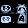
Vendors at the April International Symposium of Virtual Colonoscopy in Boston showed off a crop of increasingly sophisticated products for the VC market. Virtual colonoscopy workstations and software were the main event, but a variety of support products were also on display in the booths.
Vendors conducted training sessions for attendees on the various workstations, and each firm had a chance to present a brief overview of its products. The talks are summarized below in their order of presentation.
E-Z-EM/Vital Images
Specializing in the manufacture of contrast agents for gastrointestinal radiology, E-Z-EM celebrated its 40th birthday last month. The Westbury, NY, firm has spent eight years developing products for the virtual colonoscopy market, and now offers a wide range of bowel prep products, tagging agents, a CO2 insufflator, and a 3-D virtual colonoscopy workstation developed in cooperation with Vital Images of Plymouth, MN.
The company’s focus has always been on patient comfort, said vice president of imaging products Archie Williams. The goal was to make virtual colonoscopy so comfortable that the screening population would want to participate -- and patients would rush to tell their friends that the experience wasn't really so bad. Patients have traditionally showed up for imaging starved and exhausted from bowel prep, Williams said, so the firm wanted to improve the experience, along with the imaging results.
"We developed (NutraPrep) low-residue nutritional support that gives the patient nutritional support in the 24-hour period prior to the exam," he said. "In the late 1980s, we developed a low-sodium prep kit (LoSo Prep) to go along with the nutritional support. We developed (Tagitol) tagging agents to tag any stool retained in the colon after the nutritional laxative regimen."
The Protocol CO2 colon insufflation system keeps pressure at a uniform 25 lbs, minimizing the spikes that can occur with hand insufflation, Williams said. It adjusts automatically to equalize pressure in the event of a spasm or change in position, to enable uniform distension of the colon and a high comfort level for the patient, he said.
Vincent Argiro of Vital Images described the E-Z-EM InnerView GI workstation, which is based on Vital Images’ Vitrea 3-D viewing and workflow system. Vitrea demonstrated the first volume-rendered endoscopic simulation at the 1994 RSNA meeting, he said.
InnerView GI enables a complete zoomed axial survey of the dataset, Argiro said. Specific findings can then be noted with a marking arrow, which provides an immediate problem-solving 3-D view called virtual biopsy. This tool correlates the location of the finding. The axial and multiplanar reconstruction views can also be viewed as thick volume-rendered slabs, he said. A fly-through mode provides simultaneous updates of the endoscopic view in both forward and reverse directions.
"It's also possible, with just one click, to convert the MPR slices to transaxial and vertical and horizontal projections in the plane of the endoscopic fly-though," he said. "With one click you can slip into full problem-solving mode; with one click you can shift between the two datasets."
Vital Images has been working in the field of visualization for 15 years, and has been engaged in research even longer, Argiro said. More than 500 Vitrea systems are not only installed but are in active current use, he said. The firm doesn't work alone commercially, but instead establishes working relationships with other firms such as E-Z-EM to develop and incorporate its visualization systems.
Vital Images has also established a major R&D relationship with Japanese multimodality vendor Toshiba Medical Systems to adapt its 3-D technology for Toshiba’s multidetector CT scanners. Vital Images also recently announced a partnership with the surgical navigation division of Minneapolis-based Medtronic.
Viatronix
Viatronix president and CEO Jim Mieszala said the firm began doing work in 3-D volume rendering at the State University of New York, Stony Brook, in 1996, building gastrointestinal prototypes to perform virtual colonoscopy. However, processing power being what it was, the visualization system had to be used on what was then a million-dollar computer platform. So the company (also based in Stony Brook) decided to wait for computer processing power to catch up with its system requirements.
"It did so in about 1999, and we incorporated in 2000," Mieszala said. "Since then, we've been building our customer base and our technical base, with FDA clearances in virtual colonoscopy and calcium scoring."
Extensive development work has led to U.S. patent protection in electronic cleansing, automatic centerline, colon segmentation, "painting" of the mucosal surface to indicate areas examined, and so-called translucent rendering to distinguish stool from polyps to reduce the number of false positives, he said.
"Part of what makes us unique is the ability to do live volume rendering, and to actually fly off the (lumen) centerline," Mieszala said. "This allows the user to take a much closer look at pathology of importance," he said, and it also enables polyps to be viewed from multiple angles and under different lighting conditions.
The workstation also features template-based reporting adapted from the firm's calcium-scoring module, and offers other workflow-enhancing features based on user feedback.
"We look forward to providing advanced software that drives the acceptance of virtual colonoscopy," he said.
Philips Medical Systems
International product manager Bert Verdonck, Ph.D., said that Philips' medical imaging profile has grown considerably in the past two years following the firm's acquisition of several major medical imaging firms, including ADAC Laboratories, Agilent's Healthcare Solutions Group, and Marconi Medical Systems.
The Best, Netherlands-based vendor's primary VC tool is the Endo 3D application of Philips' EasyVision multimodality image processing workstation.
"Endo 3D is an interactive working environment which supports multiple approaches of looking into data," Verdonck said. "It supports movement tracking in various ways with reference to the original axial slices. It reformats perpendicular to the bowel through the colon," he said, and links to reference images to facilitate zooming into areas of interest.
The general volume tool enables the simultaneous comparison of prone and supine scans, or previous exams with current exams, or precontrast and postcontrast data. Unique volume-rendering techniques provide accurate surface delineation without rendering artifacts, even using single-slice CT, he said. Autobowel tracking has an automatic centerline that allows for manual guidance.
"Our most recent feature is the fold function, which allows you to look behind (haustral) folds. You can go forward and backwards, right view, left view, up and down," Verdonck said. The measurement function refers back to the original axial slices for accuracy.
Voxar
Voxar doesn't make 3-D workstations, said CEO Andrew Bissel. It makes affordable software that turns ordinary PCs into advanced workstations, though the application works on everything from PACS workstations to laptop computers.
The software delivers 2-D review of axial slices, instantaneous 3-D to confirm polyps, interactive MPR of both prone and supine studies, volumetric measurements, and the ability to easily look at two datasets at a time. The user interface tools include the ability to navigate both prone and supine datasets very quickly.
Tracking movements are supported in various ways with reference to the original axial slices, said company spokesman John Harper, who also took part in the presentation.
"The software was developed through a collaborative effort between Voxar (of Edinburgh, Scotland) and Dr. Matthew Barish and Dr. David Nicholson of the Boston Medical Center," Harper said. "So we've really written the software with radiologists' workflow in mind. The end product of this process is to produce a report as you read. Radiologists get used to taking little notes as they go along; we try to avoid that process," he said.
For example, the radiologist can select a polyp and then check a box to insert a report comment. At the end, the automated reporting process prints a completely formatted report that includes images of the areas of interest, Harper said.
GE Medical Systems
With its Advantage Navigator and associated applications, GE Medical Systems (Waukesha, WI) is developing a complete workflow strategy and toolkit the radiologist can use to look at the 2-D image, bookmark a polyp, and look at it in 3-D view and fly-through modes, said spokesperson David Spencer.
"We can look at the prone, supine, and coronal views simultaneously, all synchronized," he said. "We can bookmark the images that have a polyp, and also customize that screen. The radiologist can quickly scroll through these images, and if the patient is negative, make the diagnosis in under 5 minutes."
The average read time for positive cases is 15 minutes, Spencer said. Cases that need more attention can be viewed in the analyze phase, which visualizes 3 oblique images in addition to the endoluminal view. A novel view called virtual colon dissection opens up the colon or part of it for analysis.
There are multiple simultaneous modes of confirmation, and independent evaluation has shown that the integrated workflow design is easy to use, he said.
By Eric BarnesAuntMinnie.com staff writer
June 3, 2002
Copyright © 2002 AuntMinnie.com


