A sizable study lends new weight to the argument that virtual colonoscopy finds the riskiest colorectal polyps by nailing the largest ones. The results suggest that if VC surveillance were substituted for colonoscopy, some polypectomies of smaller lesions could be safely eliminated.
The retrospective study by researchers from the University of Wisconsin Medical School in Madison focused on adenomas, the advanced colorectal polyps considered most likely to progress to colon cancer. In previous studies, small polyp size has been shown to be a fairly reliable indicator of benign histology, although there are rare exceptions.
Researchers hope to create a rescreening algorithm that is frequent enough to catch nearly all advanced adenomas before they become cancerous, but infrequent enough to be cost-effective while encouraging compliance among the screening population.
In one colonoscopy study, 657 of 1,228 polyps smaller than 10 mm were adenomas, but neither of the two carcinomas found was smaller than 5 mm (European Journal of Surgery, October 2001, Vol. 167:10, pp. 777-81).
Adenomas come in three histologic types, each with greater malignant potential: tubular, tubulovillous, and villous. While up to 25% of adenomas may progress to cancer over time, the vast majority of polyps -- 98% to 99% -- never undergo the transformation to malignancy.
By examining the morphology, histology, and size of polyps initially detected on VC, then removed at colonoscopy, the University of Wisconsin researchers lent new evidence to the discussion of which types of lesions are being found and resected in colonoscopies of screening subjects, and whether VC can find lesions with the greatest potential for malignancy over time.
The researchers combined two large databases comprising more than 3,500 asymptomatic individuals at average risk of colorectal cancer. They studied adenomas 6 mm or larger that were removed in following virtual colonoscopy, then optical colonoscopy and polypectomy.
"Advanced adenomas are an important concept in colorectal cancer screening. They're defined as colonic adenomas 10 mm or greater, those with a villous component, or those with a focus of high-grade dysplasia," said Dr. David Kim in a presentation at the 2006 American Roentgen Ray Society (ARRS) meeting in Vancouver.
In routine colonoscopy practice, every found polyp is removed regardless of its risk of malignancy, introducing invasiveness and a small risk of complications into the task of colon cancer screening. As a result, the question of whether virtual colonoscopy can find all of the adenomas is key to determining whether the number of colonoscopies can be safely reduced.
"If we were able to delineate and extract the advanced adenomas from the larger population of polyps, this would represent a more efficient marker for polyp removal," Kim said.
The study examined the prevalence and characteristics of advanced adenomas detected at VC in a screening population, and ultimately removed at colonoscopy. The mean age was 57.3 ± 8 years, 50.3% women 49.7%, men. A total of 325 adenomas 6 mm or larger were removed at colonoscopy from the database of 3,536 subjects.
"Pathologically proven adenomas 6 mm in size or greater were extracted from the database, and from this group advanced adenomas were determined," he said.
Excluded from consideration were 119 polyps 6-9 mm removed from patients currently enrolled in a VC surveillance program, in addition to patients with annular anal carcinomas.
"We looked at the prevalence of advanced adenomas, the size, histology, morphology, and location," Kim explained.
The results showed that 124 of the 325 (38.2%) lesions were advanced adenomas, a prevalence of 3.1% in the subject cohort. The prevalence of invasive carcinoma (cancer extending beyond the muscularis mucosae) was quite low at 0.17%, as was the prevalence of high-grade dysplasia at 0.14%, according to Kim.
"The advanced adenomas in our series were large, with a mean size of 16.6 mm," he said. "The vast majority, 94.4%, were 10 mm or greater in size. There were only seven lesions in the medium-sized 6-9 mm category (comprising) 5.6% of advanced adenomas."
All invasive carcinomas were larger than 10 mm, as were four of five adenomas with high-grade dysplasia, he said.
The adenomas were classified as having tubular histology in 54.8% of the cases, tubulovillous histology (25% to 75% villous component) in 38.7%, villous histology in 4%, and serrated histology in 2.4% of the cases, Kim reported.
Morphologically, sessile, and pedunculated morphology accounted for the majority of the advanced adenomas at 45.1% and 46%, respectively. In all, 8.1% of the advanced adenomas were classified as flat lesions (height half the size of base or less), though the criteria made this characterization somewhat subjective, Kim said.
 |
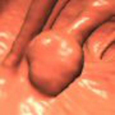
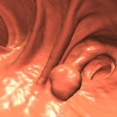
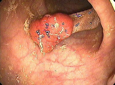
| A pedunculated polyp was shown to be a tubulovillous adenoma at histology, seen at 3D endoluminal virtual colonoscopy (above) and optical colonoscopy (below). Images courtesy of Dr. Perry Pickhardt. |
 |
Using the splenic flexure as the dividing line between distal and proximal adenomas, 42.7% were proximal and 57.3% distal. Lesions 6-9 mm in diameter represented just 5.6% of the total adenomas, or 1.4 advanced adenomas per 1,000 subjects. Nearly all medium-sized advanced adenomas had tubulovillous or villous histology; one had a focus of high-grade dysplasia. There was no evidence of invasive carcinoma among the medium-sized adenomas.
"Large size may be a useful surrogate for advanced adenomas, and ... following surveillance protocols in the medium-sized group, may be feasible given the low prevalence of high-grade dysplasia and invasive carcinoma," Kim concluded.
A member of the audience asked Kim what the gastroenterologists at his facility think about the idea of surveillance versus removal of medium-sized lesions. The practice currently offers VC subjects who present with a medium-sized polyp the option of colonoscopy or a VC surveillance program, advising patients of the risks and benefits of each decision.
"We actually had lot of resistance at the beginning, but we now have a good working relationship," Kim responded. "There's a subset (of gastroenterologists) that firmly believes in our results, and another group that has some reservations. But their feeling is that there is a tremendous number of polyps being removed in order to remove the precursor lesions to effect the incidence of colon cancer. All of this polypectomy does not decrease risk. So if we can find a way to get rid of unnecessary polypectomies without sacrificing what we're trying to do in terms of cancer risk, then I think it will be favorable, and I think the gastroenterologists are starting to see that."
By Eric Barnes
AuntMinnie.com staff writer
June 8, 2006
Related Reading
VC effective in postpolypectomy follow-up, March 4, 2006
Adverse events weigh on colonoscopy mortality equation, February 6, 2006
Ursodeoxycholic acid may help prevent colorectal adenoma recurrence, June 7, 2005
Colonoscopy performed too often after polypectomy, August 17, 2004
Copyright © 2006 AuntMinnie.com


