Some of the best ideas developed for virtual colonoscopy over the years are being refined and combined to create an advanced exam tool. Researchers from Japan are building Navi-CAD, a comprehensive VC exam system that combines unfolded views of the colonic mucosa, automated polyp detection, and unobserved region detection.
"The colon Navi-CAD system can visualize inside the colon lumen interactively and also has some (computer-aided detection) functions for detecting colonic polyps," said Kensaku Mori, Ph.D., an associate professor of information sciences from the Graduate School of Information Sciences at the University of Nagoya.
In a presentation at the 2006 Computer Assisted Radiology and Surgery (CARS) conference in Osaka, Mori said the need for better VC systems is growing in Japan, along with colorectal cancer rates, as a result of increasing adoption of Western diets in that country. The findings are corroborated, if imprecisely, by U.S. research showing that the typically low colorectal cancer rates seen in Japanese immigrants to the U.S. rise to the level of the overall U.S. population by the third generation.
As a result, early detection of colorectal disease is growing more important in Japan, Mori said. But traditional screening methods such as conventional colonoscopy have disadvantages, including a small risk of injury to the patient, as well as associated "physical and mental pain," he said.
For virtual colonoscopy, on the other hand, the tortuous colon and its maze of folds and protuberances make it a difficult target, and adequate 3D visualization tools have been lacking, according to Mori. For 3D endoluminal viewing in VC, "you have to change the viewpoint many times to see the entire colon, (and) it is also a time-consuming task; you need two to three minutes to fly through the colon," he said.
A team effort
Navi-CAD is a prototype system being researched in multiple institutions in Japan, including, among others, the University of Nagoya, Kyushu University, and Sapporo General Hospital. The system offers several features to save time and improve lesion detection, beginning with a synchronized display of several views of the colon in real-time.
Visualization modes include virtual unfolded (VU) view, which enables the display of large areas of the colonic wall; the virtual colonoscopic (3D endoluminal) view, which enables 3D viewing of the colon surface without distortion; and multiplanar reformatted (MPR) view, which enables the detection of extracolonic abnormalities, Mori said.
Finally, an "outside view" of the colon is handy for lesion localization and assessing which areas of the mucosa were missed in the first pass of the exam. "The system has a real-time visualization system, so all the analysis of the data is displayed in real-time, and in multiple views," Mori said.
The components described herein address the complex tasks required to navigate and visualize the colon.
The unfolding of CT data in the GI tract, described by a number of authors including Wang et al in 1998, is an electrical field-based method of creating a flat view of the colon. The VU view method described by Bartoli et al, and in another paper by Mori's colleague Masahiro Oda, Ph.D., generates unfolded views by controlling nonlinear ray-casting directions in the volume-rendering method, he said.
The method is based on rays cast from a central lumen path in directions perpendicular to it, Mori said. However, unfolding the colon with ray casting creates two main problems the group had to solve in its current study. First, artifacts in the form of spurious holes occur in these unfolded views, "especially in very sharp curves to the colon," he said.
The current study by Oda, Mori, and their team was presented at CARS. It employs a node-spring model to reduce the intersections of the rays, reducing the size and quantity of spurious holes that can introduce reading errors.
The correction method allocates a series of springs between planes perpendicular to the colon centerline. The plane directions are modified by the spring forces, leading to a reduction in the intersection of the planes, Oda et al explained in an accompanying abstract.
"By using the ray intersection reduction process, haustral folds and polyps were clearly distinguished in virtual unfolded views," Mori said, adding that the process "significantly reduces spurious holes." Still, the method needs further improvement, he said.
A second problem is the significant distortion in the unfolded views that occurs in narrower areas of the colon, due to the uniform height of the typical flattened colon view. The researchers reduced this distortion source by changing the height of the flattened views to correspond to the thickness of the individual segments.
The two corrective methods were applied to abdominal CT images acquired with a 512 x 512 field-of-view, 0.35-0.78 mm/pixel, 33-465 slices, slice thickness of 1-10 mm, and reconstruction intervals of 0.62-10 mm. The ray reduction process took about three minutes of computing time per case, plus an additional minute for the height-changing process.
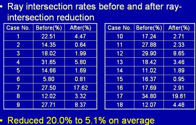
The results showed significant qualitative and quantitative improvements in the data. In the 22 cases tested so far, the average intersection rate decreased by 20% to 5.1% after applying intersection reduction, Mori said.
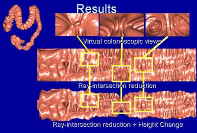 |
| Ray and height distortion reductions reduce spurious holes and visual distortion of thin areas of the colon significantly. |
 |
The prototype system also enhances the VU views by coloring them based on CT attenuation values under the colonic wall. "Because CT values of polyps tend to (be high), this visualization method is useful for the diagnosis of CT images," the researchers explained in their abstract.
Computer-aided detection
Computer-aided detection (CAD) "is a hot topic in virtual colonoscopy," Mori said, one that is increasingly important in practice for improving diagnostic performance and reducing diagnostic time.
With Navi-CAD, an automated polyp detection component is incorporated into the navigation function, he said. As the system generates fully synchronized VU, virtual colonoscopy, and CT slice views in real-time, the automatically detected polyp candidates are overlaid on them, Mori said.
Most CAD schemes use a local curvature-based method for detecting convex regions in order to extract polyp candidates. This necessitates the use of derivatives of the CT values at the vicinity of a point on the colonic wall for computing the curvature at the point. The problem with these schemes -- including the research team's previous method -- is their great sensitivity to image noise and irregularities on the colonic wall that generate large numbers of false-positive detections.
To avoid this problem, Navi-CAD substitutes the local curvature-based method for a surface-fitting method, significantly reducing the number of false positives generated, Mori said.
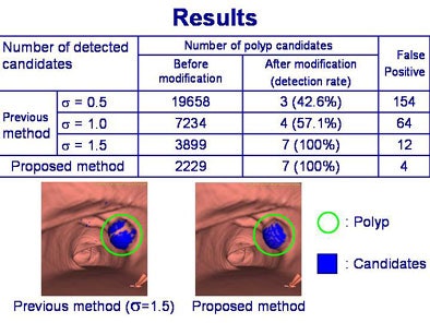 |
| Table shows CAD results in clinical data before and after incorporation of curve-fitting method. Previous method with Gaussian smoothing of σ = 1.5 was able to detect all colonic protrusions. However, false positives were too high compared to the current scheme because of false-positive detections on haustral folds. The proposed method eliminates this major source of false positives, although further development is needed. |
With their method, the inner surface of the colonic wall is fitted by a quadratic surface, from which the curvatures are calculated. "First, we calculate the derivatives along the x-, y-, and z-directions at each point on the fitted surface, and then the Gaussian and mean curvatures at the point are calculated from the derivatives," Mori said.
These curvatures are used to classify surface shapes as pits, valleys, saddles, ridges, and peaks of colonic polyps, each type color-coded for ease of reading. Regions showing the peak shapes are then selected as polyp candidates, he said.
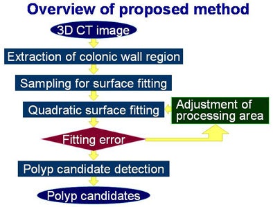 |
| The Navi-CAD's real-time CAD scheme is based on a surface-fitting method that fits quadratic surfaces to the colonic wall. The method then uses the resulting surface to compute shapes of colonic structures for identification of polyp candidate. |
The proposed CAD scheme is quite robust in the presence of image noise, Mori said. After application of a size and shape-based false-positive reduction method, the number of false positives was sharply reduced, and the detection rate for the targeted polyps in clinical data was 100% with four false positives, Mori said.
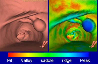 |
| Shape classification color-codes the polypoid region (right side on the image) into a shape class typically associated with colorectal polyps. |
The CAD method was initially described in a paper last year (Medical Image Computing and Computer-Assisted Intervention - MICCAI 2005, 2005, Vol. 3749: Part I, pp. 696-703).
Path tracking
The final step uses a path-tracking algorithm to first mark the entire colonic lumen as undisplayed. If a surface-rendering technique is used for the colon exam, a series of triangular patches are displayed on the screen, indicating areas covered. Alternatively, if a volume-rendering technique is chosen, voxels displayed on the screen during the exam are identified by the change in accumulated opacity values along the ray generated for the volume rendering, Mori said.
The unviewed regions are displayed on the screen and examined in a second pass through the colonic lumen. Undisplayed regions from the unfolded view are overlaid on the outside view of the colon.
 |
| A path-tracking algorithm marks undisplayed regions of the colon in blue, as shown in outside view of the colon (above) and 3D endoluminal view (below). |
 |
Using the marking system, the group found that a second pass through the colonic lumen reduced missed areas from 25% to 32% of the mucosa for a typical endoluminal fly-through, down to 6% for the endoluminal view and as low as 2.2% for the VU view after a second pass to eliminate marked areas, he said.
The Navi-CAD prototype combines a number of VC functions including unfolded, endoluminal, MPR, and outside views, along with advanced computer-aided polyp detection, and tracking of unobserved regions, Mori said.
Development plans include making the system more user-friendly. And the CAD function needs further refinement, including further reductions in false positives, Mori said. Finally, the team hopes to incorporate Navi-CAD's functions into the performance of conventional colonoscopy as well as virtual colonoscopy.
By Eric Barnes
AuntMinnie.com staff writer
August 17, 2006
Related Reading
2D primary reading plus CAD has an edge in VC study, July 11, 2006
VC software highlights colon's unseen areas, June 16, 2006
New VC reading schemes could solve old problems, October 13, 2005
CAD aids polyp detection in 64-slice study, May 12, 2006
'Filet view' VC software slices reading time, October 6, 2005
Copyright © 2006 AuntMinnie.com


