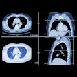
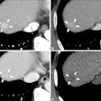
CT angiography (CTA) images are clear and readable at various heart rates on 64-detector CT, according to a new study by researchers from Switzerland. But while overall heart rates made little difference for visualizing the coronary arteries, heart rhythm mattered far more. Beta-blockers reduced artifacts across the board, but were more useful for taming irregular heart rhythms than for slowing heart rates, the group noted.
"Despite the increase in temporal resolution from four- to 16-detector-row CT, coronary CT angiography still remains sensitive to motion artifacts, which especially occur with higher heart rates," wrote Drs. Sebastian Leschka, Simon Wildermuth, Thomas Boehm, and colleagues from University Hospital Zurich in the online edition of Radiology. "The introduction of 64-section CT enables further improvement in temporal resolution and speed of volume coverage. The initial experience with this scanner type has shown a sensitivity of 94% and a specificity of 97% for the diagnosis of substantial (>50% of luminal diameter reduction) coronary artery stenoses, but the influence of heart rate on image quality was not assessed" (Radiology, September 11, 2006).
The study prospectively evaluated the effects of average heart rate and variability in heart rate on image quality using a 64-detector CT scanner (Somatom Sensation 64, Siemens Medical Solutions, Malvern, PA).
In the study, 125 patients (45 women, 80 men; mean age 59.9 years ± 12.9 standard deviation [SD]) were imaged on the 64-slice scanner, 79 of whom received beta-blockers. Data were reconstructed at 5% steps between 20% and 80% of the RR interval and transferred to a workstation (Second Wizard with InSpace 4D, Siemens Medical Solutions).
The images were acquired using a detector collimation of 32 x 0.6 mm, collimation of 64 x 0.6 mm by means of a z-flying focal spot, a gantry rotation of 370 msec, pitch of 0.24, 120 kVp, and 650-780 mAs, according to the authors. Before scanning an 80-mL bolus of iodixanol (Visipaque 320, 320 mg/mL, GE Healthcare, Chalfont St. Giles, U.K.) was injected at 5 mL/sec and controlled by bolus tracking.
"The electrocardiogram (ECG) was digitally recorded during data acquisition and was stored with the unprocessed CT dataset," the authors explained. "When automatic positioning of the R-wave indicators by the software failed, a radiologist ... manually repositioned the indicators to improve the quality of synchronization."
Only coronary artery segments 1.5 mm and larger were included in the analysis in a nod to 64-detector CT's limitations for visualizing smaller vessels, and the vessels were measured with electronic calipers. The two readers noted the presence of motion artifacts and rated image quality semiquantitatively on a five-point scale, with 1 indicating no motion artifact, 3 indicating moderate artifact, and 5 indicating a nonevaluable segment.
According to the results, average heart rate was 63.3 ± 13.1 beats per minutes (bpm), with variability of 3.2 bpm ± 2.1. A diagnostic imaging score (3 or lower) was attained in 1,821 of 1,836 segments using the reconstruction interval with the fewest motion artifacts.
"There was no correlation between mean heart rate and image quality for all segments of the right coronary and left anterior descending arteries, but there was significant correlation for left circumflex artery (r = 0.33, p < 0.05)," the authors wrote. "Heart rate variability was correlated with image quality overall (r = 0.75, p < 0.001) and for each coronary artery."
Still, heart rate variability was significantly lower in patients receiving beta-blockers (2.45 bpm ± 1.53 versus 4.29 bpm ± 2.25 (p < 0.05), and as a result, image quality was significantly better in patients receiving beta-blockers (p < 0.05). The best image quality was obtained in diastole in patients with a heart rate of less than 80 bpm and in systole with faster heart rates, the authors noted.
The small diameter and rapid movement of coronary arteries and their complex geometry make artifact-free scanning a challenge, they wrote. Motion artifact constitutes the most important challenge to coronary artery imaging. The right coronary and left circumflex are especially prone to artifact due to their proximity to the right and left atria, respectively.
Although the image quality for the left circumflex artery showed a weak dependence on heart rate in the study, there was no such correlation for the right coronary, left main and left anterior descending arteries, or for all segments together. Since the left circumflex artery follows the motion of the left ventricle, the best reconstruction level is in mid-diastole, at 50% to 70% of the RR interval, the group reported. The best reconstruction interval for the right coronary artery is in late systole or early diastole, at 30% to 60% of the RR interval, "when the right atrium is relatively motionless," they explained.
"The most important finding of our study is that the regularity of heart rate during scanning is a major determinant of image quality in coronary CT angiography," the authors wrote. "With intercycle variability in heart rate, the commonly applied relative ECG-gated image reconstruction technique (i.e., performing reconstructions at a certain percentage of the RR interval) does not generate images in exactly corresponding cardiac phases. This is because the different functions within one cardiac cycle shorten or prolong nonproportionally with different heart rates."
From an imaging standpoint, because beta-blockers were more useful for regularizing heart rate than for slowing it, a possible implication of the study may be to administer beta-blockers in patients with irregular heart rhythm, even when heart rates are normal or only mildly elevated.
"In patients with higher heart rates but regular rhythm, 64-section CT coronary angiography can be performed and diagnostic image quality can be obtained without beta-blocking medication," they wrote.
By Eric Barnes
AuntMinnie.com staff writer
September 20, 2006
Related Reading
More data will be key to CTA reimbursement, September 6, 2006
More research needed to gauge CTA's benefits, August 1, 2006
Study: 16-MDCT yields too many nonevaluable artery segments, July 26, 2006
Cardiac CTA a no-go zone for some, July 17, 2006
CT angiography detects significant coronary lesions prior to valve surgery, June 8, 2006
MRI characterizes myocardial tissue in all its states, June 2, 2006
Copyright © 2006 AuntMinnie.com

