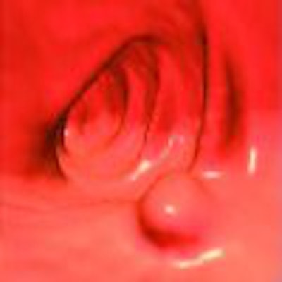
The following are several excerpts from the book Atlas of Gastrointestinal Imaging: Radiologic-Endoscopic Correlation (Saunders Elsevier, 2007), a new resource that provides information on the latest gastrointestinal imaging techniques.
Compiled by Dr. Perry J. Pickhardt, associate professor of radiology, abdominal imaging section, University of Wisconsin Medical School, Madison, WI, and Dr. Glen M. Arluk, director of advanced endoscopy, gastroenterology department, Naval Medical Center Portsmouth, Portsmouth, VA, the atlas describes the latest techniques in virtual colonoscopy, as well as all other imaging approaches for the entire GI tract.
The atlas also includes galleries of images that demonstrate the correlation between radiology and endoscopy studies to help readers see how the findings compare for every diagnosis.
Chapter 4 -- The Mesenteric Small Bowel
Often regarded as a "final frontier" owing to its relatively inaccessible location from a conventional endoscopic perspective, small bowel imaging distal to the ligament of Treitz has traditionally been the purview of the radiologist. However, the advent of capsule endoscopy and double-balloon enteroscopy has begun to shift this paradigm.
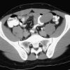
Although barium evaluation remains an important component of small bowel imaging, newer modalities, most notably CT enterography and capsule endoscopy, continue to gain importance. Perhaps the greatest strength of these studies lies in their complementary nature, because capsule endoscopy offers exquisite detail and the advanced radiologic techniques provide evaluation of the entire bowel wall and beyond.
The broad topics of small bowel neoplasia, enteritis, small bowel obstruction, mesenteric ischemia, and a variety of other pathologic conditions are covered in this chapter.
Click here to view an excerpt from this chapter.
Chapter 5 -- The Colon and Rectum
Imaging evaluation of the large intestine has undergone remarkable advances over the past decade, particularly with the advent of CT colonography (CTC, also referred to as virtual colonoscopy). Optical colonoscopy (also referred to as conventional or invasive colonoscopy) and CTC allow for complementary diagnostic evaluation of the colonic mucosa.

Optical colonoscopy remains the therapeutic gold standard for nonsurgical interventions, whereas the role of CTC in colorectal cancer screening will likely continue to expand. Cross-sectional imaging modalities such as routine CT also provide for submucosal and extracolonic investigation, including evaluation of the vermiform appendix. Although the role of the barium enema (BE) for colorectal disease continues to wane as alternative modalities evolve, its demonstration of various disease entities remains instructive.
The material presented in this chapter covers the broad topics of colorectal polyps, invasive carcinoma, colitis, diverticular disease, and the appendix, in addition to a variety of other colorectal conditions.
Click here to view an excerpt from this chapter.
Chapter 7 -- The Pancreas
Located in the anterior pararenal space of the retroperitoneum, the pancreas occupies an anatomic location that is relatively accessible by clinical examination. The combined use of CT, MR, US, ERCP, and EUS has revolutionized pancreatic evaluation.
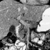
These complementary imaging modalities provide key information that guides clinical management for a diverse array of pancreatic diseases. Earlier diagnosis of pancreatitis and pancreatic ductal adenocarcinoma can lead to an improved clinical outcome.
In this chapter the clinical and imaging features of a variety of pancreatic diseases, including neoplasms, inflammatory conditions, and a host of other pancreatic conditions, are discussed.
Click here to view an excerpt from this chapter.
Copyright © 2007 by Saunders, an imprint of Elsevier, Inc.


