If you're in the market for a new premium multislice CT scanner, you'll find that picking the right system can be much more difficult than it was even a few years ago. Whereas at one time CT instrumentation developed in a relatively linear fashion -- from four to 16 to 32 to 64 slices -- vendors looking for the next big thing after 64-slice scanning are branching off into novel designs that make it harder to compare apples to apples.
A review of the state of the art in CT development finds one vendor touting a flagship system based on dual x-ray tube and detector assemblies, while another takes the linear approach even further by adding more detector rows to make a 256-slice system. Meanwhile, a third pursues enhancements to its system's imaging chain that it hopes will result in greatly improved spatial resolution for a 64-slice scanner.
Even if you don't need the bleeding edge, you'll find that vendors aren't standing still with their 64-slice technology. They are busy developing new software packages to help clinicians cut radiation dose, cope with increasingly massive datasets, and perform new applications like coronary CT angiography (CTA) and virtual colonoscopy. In the meantime, start-up companies are coming forward with highly targeted CT designs for dedicated applications like brain, breast, and ear, nose and throat imaging.
The following article is designed to help you sort through the options and make the CT purchasing decision that's right for your facility.
The rise of cardiac CT
Most healthcare facilities with more than one CT scanner use four-, eight-, or 16-slice scanners for the majority of their CT needs, but employ 64-slice or dual-source technology for sophisticated and specialized cardiac exams. Institutions that have only the most powerful scanner available must capitalize on its speed and other capabilities to improve cost-effectiveness and return on investment (ROI).
Even though cardiology studies account for only about 3% of CT procedures currently being performed in the U.S., cardiac CT captures the lion's share of attention when discussing advanced multislice CT. That's in large measure because coronary CTA gives CT -- and radiologists -- the ability to capture studies formerly conducted with catheter-based angiography -- a modality mostly controlled by cardiologists.
In terms of turf issues, the consensus of experts who spoke with AuntMinnie.com for this article suggested that those facilities that engage in active collaboration between clinicians across disciplines offer the best model for care. When colleagues cooperate, patient care -- as well as the institution -- reaps benefits. As coronary CTA gains popularity, industry leaders anticipate that the use of conventional invasive cardiac catheterization will shift into a therapeutic role in guiding percutaneous coronary interventions, such as stent placements.
Facilities pondering a CT acquisition will need to tailor the type of system they buy to the volume of cardiac imaging they think they are capable of generating. If the answer is "not much," then maybe a 64-slice system isn't necessary, and they can make do with a less powerful system.
Economic concerns
It is impossible to divorce the process of CT equipment purchasing from reimbursement issues, especially with the state of flux created by Deficit Reduction Act (DRA) of 2005, which reduced Medicare reimbursement for imaging procedures performed in physician offices and freestanding imaging centers (but not hospitals or inpatient services).
Economic issues have a huge impact on equipment utilization decisions, but the answers are more complex than you might think. While more powerful CT scanners are also more expensive, the faster patient throughput that they enable can more than compensate for their higher cost. Take a close look at the procedure volume you're currently generating -- and how much you think you could generate with a new system -- when deciding how much scanner to buy.
Many imaging facilities are also growing increasingly concerned with the vast amounts of data being generated by the new generations of CT scanners. This can impact both workflow -- how exactly do you interpret a study with hundreds or thousands of images? -- and storage costs, as multislice CT data quickly overwhelms even the most careful projections of archive usage over time.
Fortunately, vendors are developing solutions to both of these problems. As advanced visualization software grows more sophisticated with each new software iteration, many imaging facilities are shifting to 3D primary image review for more applications. New software applications are also putting new tools into the hands of radiologists, giving them the ability to analyze and interact with images in ways they never could before.
On the archiving side, a few imaging facilities have begun to develop a model of using a separate archive to store multislice CT data to prevent overwhelming a site's main PACS network. Vendors are beginning to meet this need as well with dedicated storage units that operate in parallel to the main archive.
Either way, sites looking at upgrading their CT technology need to plan for how they'll be reviewing and storing images after they get that shiny new scanner. Will the OEM's image review software be good enough, or should they go with a 3D workstation from an independent vendor? How will 3D training be handled, both for radiologists and technologists? Will it be necessary to set up a parallel miniPACS just for CT images? All food for thought.
The major OEMs
The following tracks the current product offerings from the major CT OEMs, with also a look to the future product development road map for each company.
GE Healthcare: GE's LightSpeed 64-slice platform got an upgrade at the 2006 RSNA show with the release of LightSpeed VCT XT. A centerpiece of the new model is SnapShot Pulse, a technique the Chalfont St. Giles, U.K.-based company claims reduces radiation dose by 70%, to 5-7 mSv for a typical diagnostic coronary CT study.
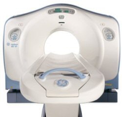
SnapShot Pulse employs a scanning technique called prospective triggered gating to ensure that x-ray exposure occurs only when the heart is at rest, in diastole. The feature is useful in two patient populations: those in whom numerous follow-up scans are anticipated, and for children with congenital cardiac anomalies, according to the company.
While the technology requires a slowed heart rate, that's not an issue when patients are treated with beta-blockers prior to scanning, according to Dr. James K. Min, assistant professor of medicine and radiology and director of the cardiac CT center at Weill Cornell Medical College and New York-Presbyterian Hospital in New York City. Nearly 90% of the hospital's patients are prepared for scanning with oral medications, therefore that factor does not negatively impact throughput.
Min's facility currently uses SnapShot Pulse in about 70% of its patients. Min described an article in press that showed a radiation dose reduction of 84% using SnapShot Pulse, which means that a typical dose is 3 mSv.
The technique can also help deal with the data explosion caused by multislice CT. "With prospective axial scanning, you literally get 300 images ... so it doesn't create large image datasets," Min said. The facility burns studies to DVD media, as well as optical disk for storage.
In future developments, GE is working on what the company calls its HD technology platform, a package of enhancements that it hopes will dramatically improve CT image quality. GE compares the new platform to upgrading to a digital camera with more megapixels that can take sharper images with each gantry rotation.
The changes include adding the ability to tune the size of the x-ray beam's focal spot to match the type of study being conducted, with smaller focal spots producing better resolution. The HD platform will also employ a new detector to resolve smaller regions of interest, and a new reconstruction engine with iterative reconstruction rather than 3D filtered back-projection reconstruction to process larger amounts of data.
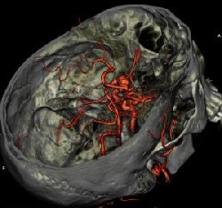
A LightSpeed system with HD should offer a number of clinical benefits. In cardiac studies, better image quality will give clinicians more confidence in determining the degree of stenosis in a coronary artery, which can have a major impact on the course of treatment. For brain imaging, the technology could help locate tiny aneurysms that might otherwise go unnoticed in the complex neural vasculature.
GE plans to showcase progress on the HD technology platform at this year's RSNA show.
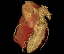
Philips Medical Systems: Brilliance 64 has been Philips' flagship CT scanner since its introduction in 2004. At the 2006 RSNA show, the Andover, MA, company introduced a prospective cardiac gating technique, called Step & Shoot, that reduces radiation dose to 3 mSv for patients with low, stable heart rates, such as those who have received beta-blockers.
Upgrading from a Brilliance 16 to the 64-slice scanner enabled increased sophistication in cardiac imaging for Dr. Samuel Wann, director of cardiovascular imaging at Wisconsin Heart Hospital in Wauwatosa. As a specialty heart hospital with five cath labs and two cardiac operating rooms in addition to a 24/7 emergency department, the hospital uses its CT scanner primarily for cardiac imaging, but about one-third of its studies are general CT. With 30% to 40% of its patients entering through the emergency department, the facility has fewer problems with reimbursement for those scans.
Wann calculates that the Step & Shoot feature delivers a radiation dose of about 3 mSv for patients, compared to 9-10 mSv for a dose-modulated cardiac CTA study and 12-15 mSv for a nondose-modulated cardiac CTA exam.
Philips has also spent considerable effort to address the workflow conundrum with 64-slice CT. The company's Extended Brilliance Workspace 3.5, introduced recently in Europe, is designed to deliver full-view and 3D volumetric images to any Philips iSite PACS or modality CT workstation, or even offsite through use of a virtual private network (VPN).
"Thin" images from the scanner are delivered to an Extended Brilliance Workspace server installed at the customer's facility. The heavy lifting and computational power are performed at the server, and the user only needs a thin-client PC with relatively modest bandwidth to read either the images or the entire clinical dataset.
Siemens Medical Solutions: Introduced at the 2005 RSNA show, Siemens' Somatom Definition CT scanner takes a unique approach to imaging beyond 64 slices -- adding a second x-ray tube and detector array to the system, enabling it to collect twice as much information in a single gantry rotation.
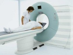
Siemens' Somatom Definition uses a dual set of x-ray tubes and detector arrays, resulting in improved temporal resolution.
The Malvern, PA, company believes the dual-source approach offers a number of advantages, including faster cardiac imaging and new capabilities enabled by dual-energy acquisition. The scanner completes a gantry rotation in 0.33 seconds, and sports temporal resolution of 83 milliseconds.
At South Jersey Radiology Associates in New Jersey, the decision to install a Definition scanner in September 2006 was driven by the system's versatility in scanning patients with varying heartbeats without the use of beta-blockers. This results in improved throughput, according to Dr. William Muhr, director of body imaging.
"With the Definition, we can scan patients with heart rates up to 120 beats per minute, so we run the scanner from 7:00 a.m. until 9:00 p.m. and run patients at four to five an hour, and I just book cardiac scans like any other CT scan," Muhr said. "It makes a difference when you can scan at normal heart rates."
Another benefit of the system is its 78-cm bore, which enables imaging of obese patients. The scanner's dual sources offer views of cardiac vessels that might otherwise be obscured in these individuals, according to the company.
For dual-energy studies, each of Definition's tubes can be set at a different energy level, which can help visualize tissues that might have different imaging characteristics based on the x-ray spectrum used. In preliminary studies in Europe, researchers have differentiated composition of kidney stones, which directs treatment decisions. In orthopedic applications, they have delineated bone from muscle from tendon tissue. Six dual-energy applications are available for clinical use on Definition.
Dual-energy imaging is also useful for vascular scans, Muhr explained. "If you have a heavily calcified plaque in the carotids or particularly below the knees, you can visualize the lumen as you would in a traditional angiogram," he said. Side-by-side images with calcium in place and "removed" via software algorithms offer important information to direct surgical intervention.
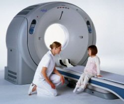
Toshiba America Medical Systems: Toshiba's flagship CT offering is Aquilion 64, which the firm began installing in 2004. But the company has made no secret of its work on a 256-slice version of Aquilion.
Rather than emphasizing the faster speed possible with Aquilion 256, Toshiba is highlighting coverage volume. The 256-slice scanner will be able to generate 128 mm of coverage per gantry rotation (256 x 0.5 mm). That means the system will be able to produce scans of complete organs without the need to stitch together images collected from multiple helical scans.
What's the clinical benefit of the volumetric approach? Toshiba researchers say it will provide major benefits in both cardiac and brain scanning. With cardiac imaging, 256-slice scanners will image the entire heart in what researchers are calling "one-beat whole-heart" imaging. Meanwhile, neurological studies will benefit from the scanner's ability to conduct whole-brain CT perfusion, subtraction CTA, and other advanced studies in a single rotation.
Just when will all this horsepower be available? Beta units have been in operation in the U.S. and Japan, and Toshiba hopes to begin shipping some time in 2008. If you can't wait that long, Toshiba has announced that the company will provide a fixed upgrade price for the 256 for those institutions that buy an Aquilion 64 scanner now.
Is a niche CT scanner right for you?
For decades CT has been dominated by multinational giants with globally recognizable names. But the last few years have seen the appearance of several smaller firms that have developed niche CT scanners for dedicated applications like brain and breast imaging. Three of these contenders are described below.
NeuroLogica
The growing utility of CT for rapid detection of neurological conditions such as stroke led to the formation of NeuroLogica of Danvers, MA. The company began selling its compact eight-slice CereTom scanner in 2006.
CereTom has a footprint that's 29 inches wide by 4 feet long, and stands 4 feet high, with the system's gantry bore at bed height. The system is capable of producing brain scans in most adults (and total body scanning of neonates), and can produce angiographic data of the head and neck in as few as 15 seconds. A primary role for the system is in acquiring routine, noncontrast scans of the brain for initial assessment of stroke or traumatic brain injury.
UCLA Medical Center in Los Angeles is using a CereTom scanner in the OR for real-time navigation and for postoperative quality assurance. Clinicians can determine whether or not intracranial hemorrhage is occurring while the entire operating team is still assembled. The system has also proved valuable in managing brain shift in the OR.
CereTom could be a good choice for older or smaller hospitals that need a CT in the emergency department for brain assessment, but that can't physically site a large whole-body scanner in their department.
CereTom also has a novel portable option, thanks to four wheelchair-sized batteries embedded in the base of the unit. The system functions cordlessly and can be moved "off the wall" for about an hour, then can be plugged into any standard outlet to be recharged. Images can be sent wirelessly.
Koning
Breast CT is still an investigational application, but several companies and research institutions are looking at the development of dedicated breast CT scanners. One of these is Koning of West Henrietta, NY.
Considered a work-in-progress, the Koning Breast CT scanner is currently involved in the U.S. Food and Drug Administration 510(k) clearance process, and clinical studies are under way. The first unit has been installed at the University of Rochester Medical Center in New York, with three more units under construction. Once clearance is obtained, the company expects to target breast imaging centers for the system.
The unit's conebeam CT (CBCT) architecture produces 0.25-mm slices through tissue in any projection, as well as 3D images. Because it features a flat-panel detector, images require additional time to reconstruct.
Xoran Technologies
Xoran of Ann Arbor, MI, offers two compact CT scanners for specialized applications. xCAT is a portable scanner based on flat-panel digital detectors designed to produce images in the neuro ICU and emergency department, and during surgeries. MiniCAT is another flat-panel-based system optimized for head and neck imaging applications that require high spatial resolution.
-- Cheryl Hall Harris, R.N.
Toshiba isn't neglecting its 64-slice platform, however. A recent focus for the vendor has been economic factors as it works to streamline workflow, with the company's "Sure" suite of applications offering more automated scanning.
Cardiac imaging has been a major focus, with applications designed to detect the most still period of the cardiac cycle for acquiring images. SurePlaque color codes calcified lesions versus soft plaques, while SureSubtraction removes bone from images.
New Mexico Heart Institute in Albuquerque began using an Aquilion 64 a year ago, and has found that in people with coronary artery disease that is not apparent with stress testing, CT scanning can be used to more accurately identify disease, according to Dr. Brendan Cavanaugh, director of cardiovascular CT. In the past year, three patients who had normal stress test/stress echo exams have died within 24 hours from a heart attack.
"If they have plaque, we put them on aggressive lifestyle changes and treat their LDL to get it to 70, which is the (American College of Cardiology) guideline for known coronary artery disease," he said. As an ancillary benefit, patient education is enhanced. It's easier to get a patient to stop smoking, change their diet, or take cholesterol-lowering medication if they can actually view their arterial plaque buildup on CT images.
Economic and regulatory factors
There is little doubt that the DRA has dampened overall sales of premium CT scanners, with reimbursement cuts of between 20% and 30% at outpatient imaging centers.
That being said, one industry spokesperson suggested that CT sales to hospitals remain strong, especially with these scanners being placed in the emergency department for rapid evaluation of various patients, including those with traumatic injuries, potential myocardial infarction patients, and stroke assessment.
Being able to justify the expense of a premium system must be considered very carefully, with a thorough ROI analysis. Most of the 64-slice scanners carry a list price of between $1.2 million and $1.4 million, plus approximately 10% to 15% of the capital acquisition cost annualized to maintain the system and a suggested 5% to 10% annually for upgrades to capitalize on some of the innovations that are just around the corner.
Siemens' dual-source CT scanner should be considered in another category, given its suggested list price of $2.8 million to $3.3 million, depending on configuration. That compares to the company's Somatom Sensation system, which carries a price tag of $1.3 million to $1.8 million.
If an institution were already fully utilizing a 16-slice scanner, and either its patient load had overwhelmed its capabilities or it saw the need for increased complex cardiac studies, upgrading to a 64-slice model would make sense. Except for extremely busy specialty cardiac centers, few can make the case to purchase a 64-slice scanner unless they can also use it for multiple other studies.
At South Jersey Radiology Associates, the facility justified the added expense of its Definition dual-source scanner by noting the benefits derived from no longer administering beta-blockers to slow patients' heart rates. This enables much more effective use of the scanner, which has become a real workhorse at the center, according to Muhr.
The future
CT is currently enjoying a renaissance that has made it one of the most exciting modalities in medical imaging. Vendors are pushing the envelope with both software and hardware designs, and new systems are on the horizon that promise to make even further improvements on 64-slice technology.
Acquiring the new generation of CT technology -- whether you go with a 64-slice system, dual-source, or one of the newer models to hit the market shortly -- is an infinitely more complex decision than buying multislice CT just a few years ago. Sites need to take a holistic approach and consider the impact that the new system will have on their archiving, workflow, and image review operations -- not to mention their capital equipment budgets.
But the benefits of premium multislice CT are legion. If they do their homework and make the right purchase, buyers will find that the technology can open new doors through new clinical applications, leading to healthier patients -- and healthier bottom lines.
By Cheryl Hall Harris, R.N.
AuntMinnie.com contributing writer
September 24, 2007
Copyright © 2007 AuntMinnie.com


