A new algorithm enables patients with arrhythmias to undergo the kind of ultralow-dose coronary CT angiography (CCTA) imaging once reserved for those with regular heartbeats. The technique paves the way for low-dose scanning in a new population of patients with suspected coronary artery disease.
Prospective electrocardiogram (ECG) triggering can substantially lower patient radiation exposure at coronary CTA compared with retrospective ECG gating, said Dr. Elisabeth Arnoldi from the Medical University of South Carolina (MUSC) in Charleston.
Of all the CT dose-reduction methods in use today, "the most profound reduction in radiation dose has been reported with the use of prospective ECG triggering, which is based on applying radiation only at predefined spaces of the RR interval," Arnoldi said in a presentation at the 2009 RSNA meeting. Although the technique offers dose savings as high as 70% to 80% compared to retrospective ECG-gated scans, "so far its application has been limited to patients with slow, regular heart rhythms," she said.
A new adaptive monitoring algorithm substantially broadens the population that might benefit from the low-dose exam. MUSC researchers tested an algorithm that enables adaptive online modulation of the ECG triggering sequence in patients with irregular heart rates, and compared its performance to invasive coronary angiography (ICA) in detecting significant coronary artery stenosis, Arnoldi said.
The algorithm, called adaptive cardio sequence (Siemens Healthcare, Malvern, PA), provides complex monitoring and scan delay functions to prospectively gated step-and-shoot coronary CTA. Specifically, it enables online detection of extra-systoles representing arrhythmia, pausing the scan acquisition and resuming it when the heart returns to normal rhythm. Thus, an imaging sequence missed for an uneven beat can be repeated at the same position if necessary.
 |
| Adaptive cardio sequence software (Siemens Healthcare, Malvern, PA) enables online detection of extra-systoles in cardiac arrhythmia, pausing the scan acquisition until the heart returns to normal sinus rhythm. Image courtesy of Dr. Joseph Schoepf and Siemens Healthcare. |
The study examined 20 patients (15 men; mean age, 61±14 years) with premature contractions who were referred for invasive angiography due to suspected coronary artery disease. Patient with atrial fibrillation were excluded, and beta-blockers were administered in patients with mean heart rates greater than 70 beats per minute.
Contrast volume and injection protocol were determined by a separate automated algorithm (P3TTM, Medrad, Warrendale, PA). After contrast administration, the patients were scanned on a dual-source CT scanner (Somatom Definition AS+, Siemens Healthcare) undergoing invasive coronary angiography later the same day.
CT scan parameters included 2 x 64 x 0.6-mm collimation, use of a z-flying focal spot technique, 0.33-sec gantry rotation time, 100-120 kV tube voltage, and 200-260 mAs tube current, adapted to the patient's body habitus, Arnoldi said.
 |
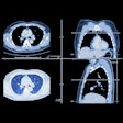
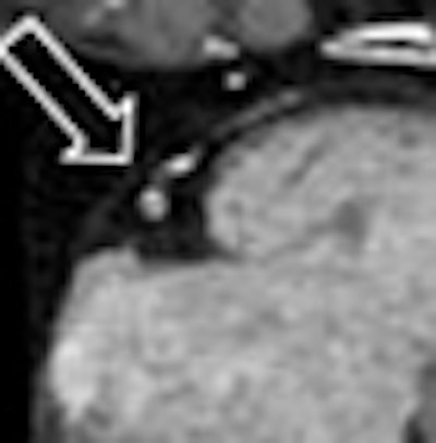
| Coronary CTA study using PT in a 57-year-old man with suspected coronary artery disease. Image at left is nondiagnostic; however, application of adaptive online modulation (right) results in diagnostic image quality. CT (above) and invasive angiography (below) show significant stenosis in the mid-right coronary artery caused by predominantly noncalcified plaque confirmed by invasive coronary angiography. Images courtesy of Dr. Joseph Schoepf and Dr. Elisabeth Arnoldi. |
 |
The adaptive modulation algorithm applied prospectively ECG-triggered radiation only during a short, predetermined time period in diastole (at 60% to 80% of the RR-interval), completely shutting for the rest of the cardiac cycle. Effective dose was calculated from the dose-length product and the conversion coefficient for the chest, i.e., 0.017 mSv x mGy-1 x cm-1.
Images were analyzed by two independent cardiac radiologists in consensus, Arnoldi said. Each coronary artery segment was examined for significant stenosis (≥ 50% luminal narrowing) using the American Heart Association's 15-segment model on a postprocessing workstation (syngo MultiModality Workplace, Siemens Healthcare).
After invasive angiography, image data were compared for the presence of significant stenosis at CCTA on both a per-segment and per-patient basis.
The researchers examined a total of 300 coronary artery segments among the 20 patients, calculating:
- Mean heart rate: 61 ± 14 bpm
- Minimum heart rate: 33 bpm
- Maximum heart rate: 83 bpm
- Mean variation of heart rate: 23 ± 19 bpm
Scan duration was 7 ± 2 heart beats using either four (n = 16) or five (n = 4) prospectively triggered image acquisitions. The mean dose-length product was 188 ± 71 mGy-cm (range, 95-311 mGy-cm), resulting in a mean effective radiation dose equivalent to 2.6 ± 1 mSv.
|
|||||||||||||||
| Results in 20 patients show high sensitivity and specificity and positive (PPV) and negative (NPV) predictive values for patients with cardiac arrhythmias. Data courtesy of Dr. Joseph Schoepf and Dr. Elisabeth Arnoldi. |
Study limitations included a small sample size, and the fact that patients with atrial fibrillation and heart rates greater than 85 bpm were excluded, Arnoldi said.
"The first initial study of prospectively gated coronary CTA in patients with cardiac arrhythmias with online ECG adaptive monitoring enables diagnosis of coronary artery stenosis with similar accuracy and comparable low radiation exposure as reported in the rest of the population," Arnoldi concluded.
Implementation of the technique in more patients and further refinement of the algorithm will broaden the range of patients who can be successfully imaged with low-dose techniques, she said.
While the algorithm in the study was developed specifically for the dual-source scanner, it should be feasible to develop similar techniques for use in single-source CT scanners, Arnoldi said.
By Eric Barnes
AuntMinnie.com staff writer
February 9, 2010
Related Reading
Studies spotlight high CT radiation dose, increased cancer risk, December 14, 2009
CAD takes on arteries with coronary stenosis detection, November 19, 2009
DECT cuts tests as a one-stop myocardial, coronary artery exam, August 10, 2009
Dual-source CTA turns in mixed results for coronary stenoses, July 30, 2009
Dual-source CT edges into cardiac SPECT turf, March 6, 2008
Copyright © 2010 AuntMinnie.com

