Routine contrast-enhanced thoracic CT exams identify unsuspected pulmonary emboli (PE) in nearly 2% of pediatric cancer patients, a prevalence level comparable to what is reported in adults.
The finding reinforces the importance of radiologists looking for this condition in pediatric cancer patients, especially in children with coagulation disorders and a history of deep vein thrombosis (DVT) or pulmonary embolism.
Pediatric radiologists from Children's Hospital Boston made this suggestion to their peers attending the recent Society for Pediatric Radiology (SPR) annual meeting held in Boston April 13-17. Their findings have also been published in the May issue of the American Journal of Roentgenology (2010, Vol. 194:5, pp. 1216-1222).
In a retrospective study to determine the prevalence, distribution, risk factors, and clinical outcome among cancer patients with unsuspected PE discovered during routine thoracic MDCT exams, two pediatric radiologists reviewed 1,002 exams performed at Children's Hospital Boston from July 2004 through April 2008. The radiologists were blinded to the specific clinical indications for the exams as well as the original radiology report findings.
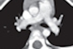
The radiologists identified nine cases of unsuspected acute PE in 468 patients, of which five had not been identified during the initial interpretation. For these five patients, PE were solitary and located either within the segmental pulmonary arteries or a lobar pulmonary artery. The researchers attributed the missed diagnoses to the relatively small size of affected pulmonary arteries and the limited extent of involvement.
Dr. Edward Y. Lee, a pediatric radiologist and assistant professor of radiology at Harvard Medical School in Boston, reported that seven of the children also had venous thromboembolism and two had tumor emboli. The pulmonary arterial locations of the 17 emboli identified were segmental (53%), lobar (29%), central (12%), and subsegmental (6%).
Three patients received thrombosis-directed treatment -- anticoagulation therapy in one case and anticoagulation therapy with embolectomy of the thrombus in two cases. One patient for whom PE was not identified received anticoagulation therapy and an inferior vena cava filter for DVT. The other patients were untreated and did not seem to experience any adverse effects, Lee said.
Related Reading
Survey: CT most popular choice for suspected PE, December 23, 2009
CT should be first approach for pulmonary emboli diagnosis, September 7, 2007
Copyright © 2010 AuntMinnie.com


