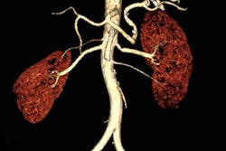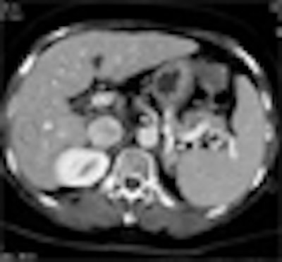
High-definition CT (HDCT) combined with adaptive statistical iterative reconstruction (ASIR) is a good way to diagnose abdominal conditions at very low radiation doses, according to researchers from Hong Kong.
The HD part is found in the 64-detector-row Discovery CT750HD scanner (GE Healthcare, Chalfont St. Giles, U.K.) introduced in 2008. Its Gemstone detector incorporates a synthetic garnet material that dissipates photon energy faster than conventional ceramics, thereby increasing speed and photon sensitivity.
In addition, the scanner's dual-energy fast kV switching registers energies faster than standard scanners, providing sharper images using less dose. HDCT can improve image quality while reducing radiation dose by as much as half for body imaging and by as much as 83% for cardiac scans, according to the manufacturer.
Once images are acquired, GE's proprietary ASIR algorithm computes low-dose image data iteratively via statistical reconstruction algorithms and algebraic reconstruction techniques that predict the characteristics of missing image data. It does this by analyzing factors ranging from adjacent image data to comparable anatomic images from a reference database.
"HDCT can provide more diagnostic information and more-consistent image quality with the ASIR algorithm," said Shen Yun from Hong Kong Sanatorium and Hospital in Hong Kong. And because ASIR effectively reduces image noise, it enables a significant reduction of radiation dose, he said.
Yun and his colleagues aimed to "evaluate the effect of ASIR on radiation dose and also image quality in CT abdomen images using high-definition computed tomography," he said in a presentation at the 2010 European Congress of Radiology (ECR).
The study included 50 consecutive patients who underwent abdominal scans on an HDCT scanner with auto-adjusted mA. Axial images were reconstructed with and without use of the ASIR algorithm.
The images were acquired using 64 x 0.625-mm collimation, 100 kV, auto-adjusted mAs, and a 22-38 noise index. ASIR was set at 30% -- meaning that the remaining 70% of the image data are reconstructed using standard filtered back projection (FBP).
So while the ASIR algorithm is fully adjustable, 100% ASIR without any FBP would "look a little bit like a painting" and isn't really practical for clinical use, Yun said, adding that some radiologists like the way the images look using ASIR settings as high as 40% or 50% ASIR.
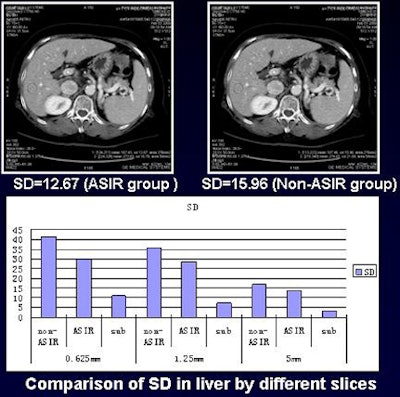 |
| Noise in the liver in the ASIR group was much lower than that of non-ASIR images, and the noise reduction was more effective for thinner slices. All images courtesy of Shen Yun. |
Noise levels (standard deviation of CT values) were measured in various regions of interest, including the liver, portal vein, bone, and kidney tissue, Yun said. The 100-kV scans are adequate for an Asian population, he said.
Two radiologists blinded to the results rated image quality in four different regions including the hepatic vein, portal vein, pancreas, and bifurcation. They used a five-point scoring system in which 1 was excellent and 5 was poor.
The average overall radiation dose was 5.19 ± 3.10 mSv, even in large-coverage scans that included the thorax and pelvis, Yun said.
The average radiation dose to the abdomen was 4.32 ± 1.92 mSv. The improvement in average image quality score (from hepatic vein, portal vein, pancreas, and bifurcation level) in the ASIR group, at 2.17 ± 0.38, was statistically significant compared to the non-ASIR group, at 2.62 ± 0.60 (p < 0.05).
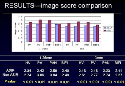 |
| Image-quality scores did not differ significantly between normal dose and lower-dose ASIR images. |
The reduction in average noise level of different tissue was also statistically significant in the ASIR group compared to the non-ASIR group. The percentage decrease in image noise of axial views was 20% in liver, 19% in vein, 9% in bone, and 15% in kidney.
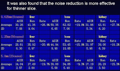
The researchers also found that noise reduction was more effective for thinner slice thicknesses.
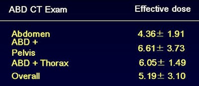 |
| The use of HDCT with ASIR enabled significant reductions in radiation dose. The average radiation dose was 5.19 mSv ± 3.10 mSv, even in large-coverage scans that included the thorax and pelvis. |
In any case, the measured noise reduction was not sufficient to describe the substantial image quality improvement obtained using ASIR, he said.
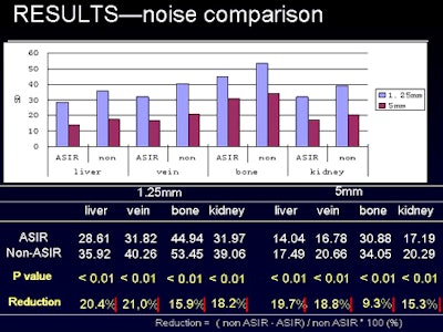 |
| Noise reduction was significantly improved with the use of ASIR (above), and noise reduction was also found to be more effective when used with thinner slices (below). |
 |
"Clearly, there is something we cannot measure using the noise index or the scoring system," Yun said. "It may have something to do with the spatial resolution or the other things involved in ASIR reconstruction, but we cannot measure that in this study."
In addition, he said, the percentage noise reduction that is obtainable depends somewhat on the quality of the original images.
"Some people claim to have 20% or 40% noise reduction -- it all depends on how noisy your original images are," Yun said. "If you have very noisy images, you will have a higher noise reduction factor" with the use of ASIR.
The study shows that HDCT can provide more diagnostic information and more consistent image quality when used in combination with the ASIR algorithm, Yun said. Second, the ability to increase the image noise when using the ASIR algorithm allows a significant reduction of radiation dose in CT abdominal imaging, he said.
By Eric Barnes
AuntMinnie.com staff writer
May 4, 2010
Related Reading
Economical DR, extremity MRI among GE ECR highlights, March 8, 2010
ASIR cuts dose in Crohn's disease patients, December 4, 2009
ASIR reconstruction sharpens images, slices abdominal CT dose, May 21, 2009
New algorithm sharpens coronary artery images, July 14, 2008
GE focuses on high-res CT with launch of LightSpeed CT750 scanner, May 13, 2008
Copyright © 2010 AuntMinnie.com


