The image quality of contrast-enhanced CT scans of the pancreas was pretty much equivalent between fast 320-detector-row contrast-enhanced scans of the pancreas and 64-detector-row scans, according to a new study in Radiology. But there were some differences, notably far lower radiation doses with the 320-row scans.
Considering that dose was reduced by nearly half when patients were scanned in 320-detector-row volume scan mode, the barely perceptible drop in image quality the researchers also noted was considered inconsequential, and unlikely to impact diagnosis, according to a group led by Dr. Satoshi Goshima from Gifu University School of Medicine in Japan. The study team also included researchers from the U.S. and other institutions in Japan (Radiology, March 15, 2011).
The authors saw potential in the wide anatomic coverage, speed, and low-dose potential afforded by the 320-detector-row scanner design. Their study aimed to prospectively compare 320-detector volumetric and 64-detector helical CT scans of the pancreas in the depiction of anatomic structures, image noise, and radiation exposure.
They examined a 154-patient cohort (85 men, 69 women; age range, 26-85 years; mean age, 67 years) who underwent biphasic contrast-enhanced CT of the pancreas. All scans were acquired on a 320-detector-row scanner (Aquilion One, Toshiba Medical Systems), operating in 320-row and 64-row modes. Patients were randomized to 320-detector volumetric scanning mode or 64-detector helical mode.
Patients were randomly assigned to one of the two scan modes for biphasic (arterial and pancreatic phase) contrast-enhanced CT.
Patients in both scan modes were scanned with 500-msec gantry rotation, using 120-kVp tube voltage and 404 mA (range, 292-500 mA) for the 320-detector-row volumetric mode and 402 mA (range, 273-500 mA) for the 64-detector-row helical mode. Both scanners acquired 16 cm of anatomic coverage: the volumetric scan in a single rotation and eight tube rotations for the 64-detector mode. Automated tube current modulation was used in the z-axis (Auto Exposure Control, Toshiba).
For review, transverse CT images were reconstructed at a section thickness of 0.5 mm, with no overlap in either group. Arterial-phase volume-rendered CT angiograms, arterial-phase multiplanar reformatted (MPR) images, and pancreatic parenchymal phase MPR images were created by a trained technologist using a workstation (Advantage Windows 4.2, GE Healthcare).
Two experienced radiologists working independently reviewed the arterial- and pancreatic-phase transaxial images, arterial-phase CT angiograms, and arterial- and pancreatic-phase MPR images in five sessions, each separated by at least two weeks and with random case presentation, the authors wrote.
The results showed no significant differences between the two groups in HU values in the abdominal aorta or pancreas, with p values ranging from 0.15 to 0.78.
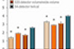
However, the radiation dose was far lower in the 320-detector-row volumetric scans. Mean dose-length product (DLP) was 43% lower in the 320-detector-row group (675.4 mGy-cm) compared to the 64-detector-row group (1187.8 mGy-cm) (p < 0.001).
The signal-to-noise (S/N) ratio of the abdominal aorta, pancreas, and abdominal wall fat on biphasic images was significantly lower in the 320-detector-row group than in the 64-detector-row group (p < 0.001) Image quality was acceptable in both groups and was slightly better in the 64-detector group for pancreatic phase axial images (p = 0.02) and arterial-phase multiplanar reformatted images (p < 0.01).
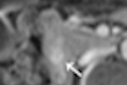
The overall image quality was equivalent, and no significant differences were found in the depiction of pancreatic parenchyma, the main pancreatic duct, focal pancreatic lesions, or splanchnic arteries.
"On the basis of quantitative analysis of pancreatic CT attenuation, the 320- and 64-detector groups in our study protocol are equally acceptable for delineation of pancreatic and adjacent visceral structures during pancreatic phase imaging," Goshima and his team wrote.
The 43% reduction in DLP at 320-row CT "may be attributable to the fact that the large cone angle used in the 320-detector scanner leads to a smaller contribution of overbeaming to the total dose, as well as to the fact that there was no overranging, as seen in helical acquisitions," they wrote.
A previous study by Siebert et al found 64-detector-row scanning to be superior for vessel depiction, particularly small vessels, while the present study found no significant differences.
"Only a few small arteries, such as the dorsal pancreatic artery and peripheral branches of the left gastric artery, were less well-visualized in the 320-detector group," Goshima et al wrote of the present study, positing that the pronounced artifacts at the skull convexity may have inhibited visualization in the earlier study by Siebert and colleagues.
As for the lower S/N ratios in volumetric mode, "The 320-detector scanner with a large dimension of detector in z-axis may require a broad cone angle; thus, the resulting scattered radiation is likely to increase and cause image degradation," they wrote. "Another reason for image degradation with 320-detector scanning can be the lower photon flux in the 0.5-second acquisition with volumetric scanning compared with the 4.2-second acquisition with helical scanning."
Even though the results show that the S/N ratios in the aorta, pancreas, and abdominal wall fat tissue and the qualitative image quality of arterial-phase transaxial images, arterial-phase MPR images, and pancreatic-phase transaxial images were significantly lower in the 320-detector group, the depiction of anatomic structures was "almost comparable" between the two groups in a qualitative assessment.
The main study limitations were that different scan modes were used in different patients in the clinical study -- and that diagnostic performance could not be compared because the different patients had different pathologies scanned only once, the group noted.
"A 320-detector CT scanner in the 320-detector volume scan mode facilitates fast volumetric contrast-enhanced CT of the pancreas, with image quality similar to that with a 64-detector helical scanner but with reduced radiation dose," they concluded.


