Monday, November 28 | 3:30 p.m.-3:40 p.m. | SSE05-04 | Room S404CD
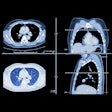
Quantifying perfusion defects at dual-energy CT (DECT) helps identify pulmonary embolism (PE) patients at risk of adverse outcomes and should be part of their risk assessment, say investigators from the University of Munich.The study, by Dr. Paul Apfaltrer and colleagues, examined the correlation of perfusion defect volume (PDvol) at DECT with CT pulmonary angiography (CTPA) obstruction scores, CT parameters of right ventricular dysfunction (RVD), and 60-day clinical outcome in patients with acute PE.
PDvol showed only a weak correlation with right ventricular/left ventricular diameter ratios in the four-chamber view, while no correlation was observed between PDvol and the remainder of the RVD CT parameters.
"In our study, of all evaluated CT parameters, PDvol demonstrated the highest predictive power for the detection of adverse clinical events," Apfaltrer told AuntMinnie.com.
Ten of the 60 patients with PE had adverse clinical outcomes, including required mechanical ventilation, catecholamine therapy, or death (n = 3).
The differences between these findings and those of previous studies may be attributable to the fact that the researchers used semiautomatic quantification of perfusion defect volume rather than a subjective score considering the number of affected segments, Apfaltrer said.
The weak correlation of PDvol to CTPA obstruction scores may have been due to "preserved perfusion in arterial segments in which there is partial occlusion above the segmental artery, resulting in areas of mismatch with partial occlusion yet normal perfusion," he stated.
Nevertheless, volumetric quantification of perfusion defects at DECT identifies patients at higher risk of adverse clinical outcome. The quantification of perfusion changes "might in the future be integrated in the risk assessment of patients with the diagnosis of PE at dual-energy CT angiography," he concluded.


