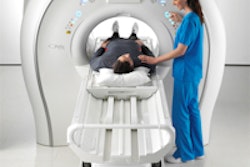
At the 2011 RSNA meeting, Hitachi Medical Systems America raised eyebrows with the launch of Echelon Oval, a 1.5-tesla MRI scanner with an oval-shaped magnet. This year, Hitachi extends the oval concept to 3 tesla with Trillium Oval, a work-in-progress scanner not yet for sale in the U.S.
The idea behind an oval-shaped bore is simple. While closed-bore magnets typically have cylindrical bores, this design hardly matches the shape of the typical patient's body habitus. As a result, cylindrical magnet bores can contribute to patient claustrophobia, a major factor behind the large number of MRI scans that are started but not completed each year.
With Echelon Oval, Hitachi created a bore shape that it believes is a better match for patients. The 1.5-tesla version of the system launched at last year's RSNA sports a bore that's 74-cm wide, compared to the 60-cm bore traditionally found on most 1.5-tesla magnets and the 70-cm bore on the newer generation of wide-bore magnets. The first installation of the system was made in the U.S. in September.
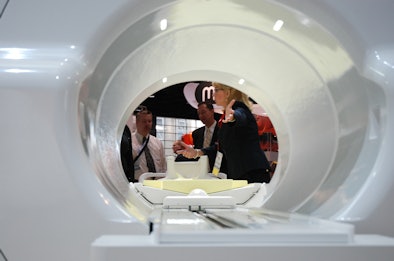 |
| Hitachi's 1.5-tesla Echelon Oval MRI. |
Trillium Oval moves the oval concept into the 3-tesla segment, which is becoming the standard for certain types of clinical imaging. The 3-tesla version also has a 74-cm bore, and like the 1.5-tesla version maintains Hitachi's 50-cm field-of-view in all directions. Hitachi is also touting the high magnet homogeneity specifications found on its scanners.
Other patient-friendly features of the new scanner include a 63-cm-wide mobile table with a 550-lb weight capacity and retractable arm boards for patient safety. Another feature designed to minimize claustrophobia is Hitachi's feet-first imaging concept, which is standard for all procedures.
Another feature that offers workflow benefits is the company's Workflow Integrated Technology (WIT) radiofrequency (RF) coils built into the table -- the feet-first concept enables MRI technologists to put 8-channel and 12-channel WIT coils into the table and never remove them except for breast procedures. Hitachi also has a spine coil that can be wrapped around the patient, leading to a 20% increase in signal-to-noise ratio.
Other features on the scanner include Hitachi's AutoPose automated slice planning technique for acquiring slice locations in the exact area with each study; isoFSE (fast-spin echo) isocentric imaging protocol, and Radar motion-compensation technique, which can be combined with the vendor's Rapid parallel-imaging protocol for studies such as free-breathing abdominal exams.
Trillium Oval is not yet for sale in the U.S. and is being shown at RSNA as a work-in-progress.
Hitachi is also rolling out new capabilities for other scanners, such as the addition of arterial spin labeling to its Veins and Arteries Sans Contrast (VASC) technique, and also the addition of FSE to VASC. Both techniques are first being launched for the Oval scanners and will be migrated to the company's Oasis open MRI magnet.
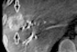
Meanwhile, Micro TE is an advanced musculoskeletal imaging protocol that offers better visualization of tendons and ligaments, and new applications in shoulder and spine imaging will be added. BeamSat TOF (time of flight) offers selective saturation of the circle of Willis.
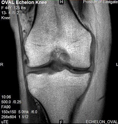 |
| Micro TE sequence (0.25-msec in this example) allows enhanced visualization of structures with very short T2 relaxation times (cartilage, tendons, ligaments) that cannot be seen with conventional imaging. |
Finally, Hitachi will show an upgrade for the Oasis called Oasis XP Workflow Enhancement. The upgrade brings new capabilities to Oasis scanners installed in the field, such as AutoPose, isoFSE, and T2 relaxation maps.
Focus on CT
Hitachi has historically been known as an MRI vendor, but several years ago the company entered the CT market, and it's making CT a significant focus of its RSNA 2012 exhibit. A major highlight is an upgrade for the Scenaria 64-detector-row scanner called Advanced 128 that offers 128-slice reconstruction.
Most CT systems from competing firms offer double-slice reconstruction; with Advanced 128, Hitachi will now offer similar technology on Scenaria, which has a 40-mm detector area with 64 channels. The company has submitted a 510(k) application to the U.S. Food and Drug Administration (FDA) for Advanced 128.
Other new CT technologies include the company's second-generation iterative reconstruction algorithm, called Intelli IP (Advanced) -- a new version of its conventional Intelli IP. Intelli IP (Advanced) features an updated reconstruction engine that doubles the speed of reconstruction from 18 images per second to 35. Intelli IP (Advanced) will also be available for Scenaria systems in the field.
Hitachi is promoting the concept that Scenaria is a scalable platform, with models starting from a basic 64-slice scanner listing less than $600,000 to an advanced 128-slice system well-suited for cardiac imaging. The company is positioning the basic Scenaria model as an option for hospitals currently using 16-slice technology.
Also new in CT are several software packages, including a simplified dose reporting feature called Simplified Dose Report that enables Scenaria users to send data to a dose registry, with the dose report embedded in the images.
Another new technology is Exam-Split, which enables users to take a long-coverage exam, such as from the emergency department, and split it into different studies based on patient anatomy to send to different clinical specialists.
Finally, Hitachi is touting Scenaria's open design, with a 70-cm gantry and 88-cm gantry thickness. The scanner's table supports patients weighing more than 500 lb, and it features a laterally shifting design called IntelliCenter that enables users to shift the table left or right by up to 80 mm. This allows the field-of-view to be focused on the area of interest, enabling the use of smaller bow-tie filters and thus less radiation dose.


