In patients with suspected pulmonary embolism (PE), a CT pulmonary angiography (CTPA) protocol using high-pitch dual-source electrocardiogram (ECG)-synchronized helical scanning outperformed standard pulmonary CTA by almost every measure, according to an article in the American Journal of Roentgenology.
In 55 patients assigned randomly to one protocol or the other, the high-pitch technique showed significantly higher signal intensity (SI) than standard CTPA, the authors wrote. High-pitch scanning also improved image-quality scores in assessing cardiovascular structures, minimizing motion in the central arteries and enhancing the pulmonary arteries. The standard pulmonary CTA technique was judged better only with images of the lung parenchyma.
"High-pitch ECG-synchronized pulmonary CTA provides higher pulmonary arterial SI, decreased motion of central pulmonary arteries, and improved assessment of cardiovascular structures with similar contrast dose and lower radiation compared with standard pulmonary CTA," wrote Dr. Michael Bolen, Dr. Rahul Renapurkar, Dr. Sandra Halliburton, and colleagues from the Cleveland Clinic (AJR, November 2013, Vol. 201:5, pp. 971-976).
Ruling out PE
Pulmonary embolism is an important problem in the U.S. with more than 600,000 cases a year, and CTPA is the gold standard imaging modality due to its ability to noninvasively diagnose or rule out suspected PE with high accuracy, the authors wrote.
For examining suspected PE, second-generation dual-source CT scanners wield advantages in their ability to operate at a pitch of 3.0 or greater using the second x-ray source and detector array to independently acquire gap-free data with a slightly limited field-of-view, according to the authors.
High-pitch acquisition using data collected from each array during one-quarter of the gantry yields high temporal resolution of 75 msec. This technique combined with ECG synchronization -- not used in conventional CTPA -- offers rapid imaging of data at a specified phase of the cardiac cycle and at very low radiation doses, the group noted.
Conventional CTPA acquired without ECG synchronization can suffer from motion artifacts from the heart and vasculature, Bolen and colleagues wrote.
The researchers prospectively examined 47 patients with suspected pulmonary embolism (60% women; mean age, 57 ± 14 years) who were randomized to either high-pitch ECG-gated CTPA (n = 26) on a dual-source CT scanner (Somatom Definition Flash, Siemens Healthcare) or the conventional version (n = 21) on a 64-detector-row single-source scanner (Somatom Sensation 64, Siemens). Bolus timing was used to ensure optimal enhancement of the contrast media.
Between the two groups, there were no significant differences in age, weight, or heart rate, and a similar amount of contrast was used, the study authors wrote. Contrast administration was in the range of 4.2 to 6.0 mL per second for high-pitch scanning and 3.8 to 5.5 mL per second for the group getting the standard-pitch protocol.
Two independent readers interpreted subjective image quality of the pulmonary arteries, as well as cardiovascular structures and pulmonary parenchyma. They measured signal intensity in one segmental artery and three central pulmonary arteries for each patient.
PE was diagnosed in 12 (26%) of the 47 patients -- with high-pitch CT showing evidence of PE in seven (26%) of 26 patients and conventional CTPA showing evidence of PE in five (24%) of 21 of patients.
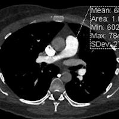
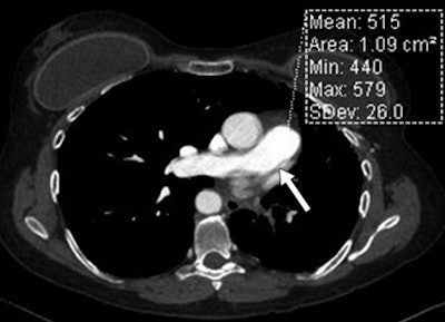
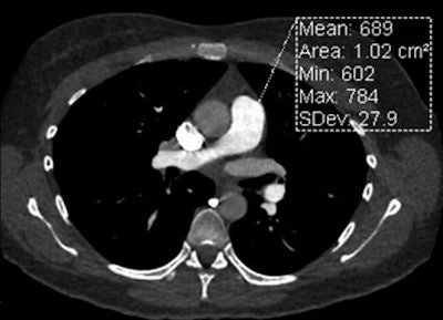
 Standard CTPA in a 50-year-old woman (above) and high-pitch ECG-synchronized CTPA in a 53-year-old woman (below). Both patients weighed 79 kg. Signal intensity is represented by attenuation values (in mean HU) in main pulmonary artery. Motion artifact of the central pulmonary artery is noted in the standard CTA image above (arrow). Images republished with permission from AJR, November 2013, Vol. 201:5, pp. 971-976.

High-pitch CTPA showed higher signal intensity for the pulmonary arteries, and image quality scores demonstrated the technique's superiority for the assessment of cardiovascular structures (p < 0.001), particularly the ability to minimize motion in the central pulmonary arteries (p < 0.001) and deliver greater pulmonary enhancement (p = 0.001). And high pitch did all of this using a significantly lower radiation dose: a mean 2.9 ± 1.4 mSv for high-pitch scanning versus 6.1 ± 2.9 mSv (p < 0.001) for conventional CTPA.
Only image quality assessment of the lung parenchyma was lower for high-pitch CT, with 8% of high-pitch cases receiving an image quality score of four (on a scale of 1 to 4, with four being best), compared with 62% of the conventional CTPA cases.
Bolen and colleagues concluded that high-pitch ECG-gated CTPA "provides higher pulmonary arterial SI, decreased motion of central pulmonary arteries, and improved assessment of cardiovascular structures with similar amounts of administered contrast agent yet lower radiation dose when compared with standard pulmonary CTA."
"Although lung imaging quality scores were higher with the standard pulmonary CTA approach, diagnostic-quality images without significant limitation were obtained for all high-pitch ECG-synchronized helical CTA cases," they wrote.
New use for high-pitch scanning
Except for the slight reduction in image quality in the lung parenchyma, the high-pitch ECG-gated acquisition technique -- FLASH for short -- had very few drawbacks, even though the study was technically an off-label use of the FLASH technique, said Halliburton, a professor of radiology at the Cleveland Clinic Foundation, in an interview with AuntMinnie.com.
"We excluded patients over 125 kg, so as long as you're restricting the size of your patients you should be OK," she said. "That's the limitation, generally speaking, of the FLASH technique: You only have so much tube output that you can manage to achieve with the superfast table feed."
The other standard limitation of FLASH is the heart rate restriction. "For coronary imaging, you can only use the FLASH technique if they have a stable heart rate, less than 60 or 65 beats per minute; otherwise, you wouldn't be able to scan the whole heart," she said.
According to Halliburton, the advantages of gating CTPA exams are at least as important as the dose savings.
"Our discussion has focused on the dose aspect, but even more significant, we think, are the advantages you have with decreased motion of the central pulmonary arteries," she said.
As for limitations, the study was aimed at quantitative and qualitative image criteria, and it did not evaluate the sensitivity or specificity of PE identification compared to a reference standard, the authors noted. Also, the subjects were all outpatients and the results may not be applicable to inpatients. Finally, the high-pitch ECG-gated technology is not widely available, limiting its potential use.


