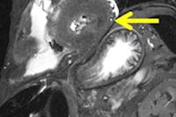
VIENNA - A ground-breaking study examining the reliability of MRI to image experimentally produced stab wounds in amputated human limbs was presented for the first time during the opening day of ECR 2014.
The Italian study involves collaboration between the University of Padua's radiology and forensic departments. It's exciting news for the growing number of radiologists imaging non-life-threatening wounds that don't require immediate recourse to CT, but that do need careful clinical management and from which images may end up as forensic evidence in criminal cases or being used in postmortem reports.
 Dr. Chiara Giraudo from Padua, Italy.
The study is the first paving stone on the path to more MR studies on stab wound patients in the routine clinical setting, whether for defining the length and direction of the wound or for diagnosis of vessel involvement for clinical management, such as vascular surgery or postmortem reporting. Little data exist about CT features of human stab wounds in the literature and until now, there were virtually no data at all about MR features of the same.
The Italian study aimed to see whether MRI could provide even more information than CT. Although not as fast as the latter, the advantage of MRI is that it can be used without contrast media, unlike its study rival, minimizing invasiveness to patients.
The researchers used both 64-slice CT and 1.5-tesla MRI to ascertain the incision length of 10 wounds, each of which was between 4 cm and 6 cm deep. CT and MRI examinations were performed before stabbing, and with the knife inside the wound track, the latter with CT only. Both MR and CT were also carried out before and after filling each wound with contrast medium. The length of the ceramic blade used to penetrate the limb was then compared with the length of wound filled with contrast medium.
Both modalities underestimated the length of the wounds inflicted, possibly due to tissue retraction once the knife was pulled out of the limb; CT underestimated wound length by an average of 11.9%, and MRI by an average of 9.1%. Although this is not a great statistical difference, and one cannot yet extrapolate from this that MRI is superior, the results generally reflect good signal with MRI and may lead to even more positive results in future, according to Dr. Chiara Giraudo, a resident radiologist at Padua who specializes in forensic and musculoskeletal imaging, and who presented the study at Thursday's scientific session on emergency radiology.
She said the limits of the study consist of the small number of stab wounds studied, their orientation and site (stab wounds may be multidirectional and have different features in different organs), and the fact that these wounds were made on amputated limbs and the results therefore don't factor in bleeding and tissue elasticity. Nevertheless, the preliminary results are promising.
"So far both CT and MRI can provide consistent and reproducible data on the injury depth," Giraudo told ECR delegates.
 Dr. Matteo Brambati from Milan, Italy.
She noted that further trials on live subjects are scheduled for next year, and will help overcome the current limitations of this study, in tandem with the creation of a new forensics unit at the hospital. Such studies are needed to confirm and explore the reliability of MRI and are due to include different MR sequences used to explore a range of wounds in various anatomical areas, not just the limbs, inflicted by different weapons. Furthermore the studies should also include a protocol for MRI without contrast medium.
Another Italian study took center stage during Thursday's session, this one also pertaining to patient management but focusing on the clinical impact of chest and abdominal CT on emergency patients. The presenter was Dr. Matteo Brambati, a radiologist at San Donate Milanese Hospital in Milan.
While the number of CT scans has increased by 20% in the past 20 years, CT only yields expected findings in 40% of cases. The study aimed to determine how CT changed clinical management of patients and also measure therapeutic variation related to CT results in each subject, he said.
Originally published in ECR Today on 7 March 2014.
Copyright © 2014 European Society of Radiology


