Tuesday, December 1 | 11:10 a.m.-11:20 a.m. | SSG03-05 | Room S404CD
In this study, researchers from Boston investigated the characteristics of lung abnormalities seen on material decomposition images from dual-energy CT (DECT).Different kV levels for each x-ray tube on dual-source CT scanners can be used to distinguish elements such as iodine and calcium, which are more attenuating than water at low kV levels.
As a result, the differential interactions of two x-ray energies form the physical basis for decomposition of images into two materials such as water and calcium, calcium and iodine, or iodine and water, wrote lead author Dr. Alexi Otrakji, from Massachusetts General Hospital, in an email to AuntMinnie.com.
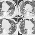
In their study, the researchers analyzed 83 scans from 83 patients on both single- and dual-source CT to determine the value of material decomposition.
"We found that evaluations of material decomposition images are useful for differentiating pulmonary opacities such as pulmonary infarcts, pneumonia, and atelectasis," Otrakji wrote.
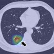
The investigators looked at pulmonary blood volume images for the presence and size of blood volume abnormalities in areas where abnormalities had been seen on other images. In cases of pulmonary embolism with infarction, decreased perfusion on pulmonary blood volume images was at least as large as pulmonary opacities.
Decreased pulmonary blood volume was also seen in six patients with nonocclusive pulmonary embolism. In patients with atelectasis, pneumonia, and emphysema, perfusion abnormalities on pulmonary blood volume images were smaller or equal to the opacities or lucencies seen on other images, revealing no size mismatch.
